Week 4: Protista
Objectives
- Define the term “protist” and explain why this is not a monophyletic group.
- Identify representatives from each supergroup Excavata, ”SAR” clade, Archaeplastida, and Unikonta
- Draw a phylogenetic tree for the eukaryotes and explain why the eukaryote supergroups form a polytomy.
- Indicate the position of plants, animals, and fungi on the eukaryote tree, and identify the group of protists most closely related to each.
- Give examples of protist species from each eukaryote supergroup.
- Give two examples of the significant impact of specific protists on their ecosystems.
After an introduction to protists, this lab includes a series of activities, each focused on one of the four Eukaryotic supergroups. The lab concludes with a set of post-lab activities that give an overview of this group of organisms.
I. What Are Protists?
Your instructor may request that you review “Reading: Protists.”
Protists are a diverse collection of eukaryotic organisms, including both unicellular and multicellular species, and are found free-living or parasitic in all kinds of habitats. The cells are complexly organized with organelles and the nuclear envelope membranes. Most protists possess a single nucleus, but some species may have two or more. They usually reproduce asexually, but sexual reproductive processes are also known to occur between two different mating types mt+ and mt-. Protists display certain characteristics that categorize them based on the organelle structure and locomotion (motility). Amoeboid use cytoplasmic projections called pseudopodia; flagellates use flagella; ciliates use cilia, or sporozoans if they lack any motility structures.
Understanding the complex history of protists and their evolutionary process is essential for our scientific discourse and discovery. In this part of the lab, we will analyze different protists to identify characteristics correlated with the cell-structure and movement. But there is little else that unifies this group—it is a term used to designate all eukaryotes that are not classified in one of the monophyletic eukaryote kingdoms. You might ask why biologists have not created monophyletic groups for the protists. This is in fact, the main question that this lab exercise will focus on. As you work through the activities, you will discover for yourself the reasons!
First, watch the videos below on the pseudopodia movement, the Euglena flagella movement, and the cilia movement in Paramecium.
From your previous courses, list as many characteristics of eukaryotes as you can.
Key Terms
| Term | Definition |
| Supergroup | An informal taxonomic level used to group organisms whose phylogeny is uncertain. For example, the eukaryotes are grouped into supergroups at a level that falls between domain and kingdom. |
| Clade | A monophyletic group at any taxonomic level (domain or kingdom, but also genus, species, or any other level) |
| Taxon | A general term use to designate a group at any taxonomic level (plural: taxa) |
| Protist | An informal name for any eukaryote that is not a plant, animal, or fungus. Does not imply monophyly. |
| Eukaryote | A microscopic single-celled or multicellular organism that has a nucleus and specialized organelles. Eukaryotes include plants, animals, and fungi. |
| Algae | A group of prokaryotic and eukaryotic organisms, usually composed of a single cell or a colony of similar cells and includes the seaweeds. |
Classification and Phylogeny of Protists
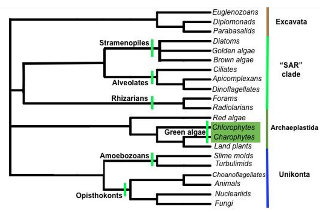
The reconstruction of evolution and phylogenetic relationships within the four Eukaryotic supergroups is still an ongoing process. With the new Whole Genome Sequencing (WGS) technologies along with the sequencing of the complete 18S rRNA markers, new members are being added to the supergroups. Not all of the clades within the supergroups are monophyletic, and an example of it is the Stramenopiles. Most of the members have arisen from the primary and secondary endosymbiosis, highlighting the fusion of the photosynthetic bacteria and heterotrophic eukaryotes. The following figure summarizes the phylogeny of Protists depicting the Four Eukaryotic Supergroups (Figure 1).
II. Supergroup Excavata
This supergroup consists of several major groups, such as: Diplomonads and Parabasalids, with modified mitochondria, and Euglenozoans including Kinetoplastids and Euglenids, containing spiral or rod inside flagella. Some of the examples of the Diplomonads and Parabasalids include Giardia and Trichomonas. Examples of Kinetoplastids and Euglenids include Trypanosoma and Euglena (see Figure 2).
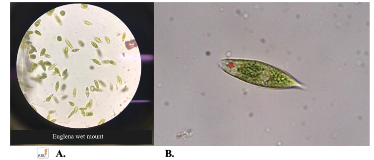
Common flagellated protozoans are found in nutrient-rich freshwater, except for a few marine species. The cells vary in length from around 20 to 300 μm, and are typically cylindrical, oval, pear or spindle-shaped with a single emergent flagellum for movement. There are usually many bright green chloroplasts, although some species are colorless. If sunlight is not available, it can absorb nutrients from decayed organic material. Euglena is also found in sewage systems. Thus, Euglena is unique like a plant carrying out photosynthesis and like an animal has a whippy flagellum to move through the water and survive on decaying matter. Euglena is a flagellated cell with the same chlorophylls (a and b) as found in higher plants. Euglena is unusual for an alga in that it can lose its chloroplasts when kept in the dark or treated with the antibiotic streptomycin, but it is able to survive as a heterotroph. Thus, this organism has an affinity for both algae and protozoa. For this reason, Euglena is claimed by botanists as an alga, but as a protozoan by zoologists.
Activity 1: Excavata Survey
View the euglenozoan specimens available and then answer the following questions. (The following questions are from the sources attributed below.)
1. What color is the euglena?
2. What structure does the euglena use to move?
3. Can you see any internal chloroplasts?
4. Can you see the red eyespot? It does not give the organism vision, rather allows it to sense the presence of light.
Trypanosoma is another example of flagellated Euglenozoan. Trichomonas vaginalis is an example causing vaginal infection and more potent trichomoniasis. Trypanosomes are microscopic, one-celled protozoans of the genus Trypanosoma, of which hundreds of species are known. A trypanosome is long, pointed and possesses a flagellum. The flagellum arises at the front, or anterior end of the parasite and curves back to form the edge of a long undulating membrane used in locomotion.
T. gambiense and T. rhodesiense cause African sleeping sickness, and both are transmitted by tsetse flies. Learn more about this disease by watching the video below, and then answer the questions that follow. (The following questions are from the sources attributed below.)
1. What part of the human body does the Trypanosoma invade?
2. What structure does the Trypanosoma use to move?
3. How does the Trypanosoma avoid being killed by the white blood cells?
4. Can African sleeping sickness cause death?
III. Supergroup “SAR” Clade (Stramenopiles, Alveolates & Rhizarians)
This supergroup consists of several major groups, such as: Stramenopiles with hairy and smooth flagella, Alveolates containing membrane-enclosed sacs (alveoli) beneath the plasma membrane, and Rhizarians with threadlike pseudopodia. Some of the examples of the Stramenopiles include diatoms, golden and brown algae and Trichomonas. Examples of the Alveolates include dinoflagellates, Apicomplexans and Ciliates. Examples of the Rhizarians include Foraminiferans and Cercozoans.
Stramenopiles
The Stramenopiles include diatoms. Diatoms belong to a large group called the heterokonts, including both autotrophs (e.g. golden algae, kelp) and heterotrophs (e.g. water molds). There are more than 200 genera of living diatoms, and it is estimated that there are approximately 100,000 extant species. Diatoms have two hard cell walls (called frustules) composed of silicon oxide. Their yellowish-brown chloroplasts contain pigments such as fucoxanthin. Diatoms are a widespread group and can be found in the oceans, in freshwater, in soils, and on damp surfaces. Most live in open water, although some live as surface films at the water-sediment interface, or even under damp atmospheric conditions. They are especially important in oceans, where they are estimated to contribute up to 45% of the total oceanic primary production.

Activity 2: Stramenopile Observations
Answer the following questions. (The following questions are from the sources attributed below.)
1. View the diatom specimens available and then answer the questions below.
a. What material is found in the cell wall of the diatoms?
b. Are the organisms single or multicellular?
2. View the brown algae specimens available and then answer the questions below.
a. What pigment does brown algae use for photosynthesis?
b. Name and describe the characteristics of one brown algae specimen.
3. View the dinoflagellate specimens available and then answer the questions below. Note that some experts classify the dinoflagellates as a sister group to the rest of the stramenopiles, rather than as part of the stramenopiles. This demonstrates the complex and ongoing investigations into protist phylogeny.
a. What structure does the dinoflagellate use for movement? How many of these structures does it have?
b. Are the organisms single or multicellular?
Alveolates
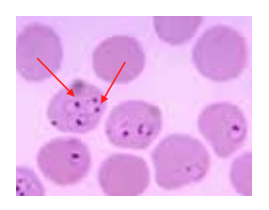
Members of this group are referred to as sporozoans as they lack locomotion structures. An example is Plasmodium, the genus responsible for malaria (caused by Plasmodium falciparum transmitted via a female Anopheles mosquito vector) in humans and other animals. In humans, the parasite is found intracellularly in red blood cells and is used as a diagnosis of malaria. Another member of this group includes ciliated protozoan Ciliophora which possesses numerous cilia for locomotion. And Paramecium is a unicellular organism found in freshwater throughout the world. It has a stiff outer covering that gives it a permanent slipper shape. It swims rapidly by coordinated wavelike beats of its many cilia: short, hair-like projections of the cell. The paramecium has an external oral groove lined with cilia leading to a mouth pore and gullet; food is digested in food vacuoles. Paramecium can divide asexually by cell division called “binary fission.”
Tetrahymena is a ciliated protozoan with an oral apparatus used for feeding on bacteria. The organism swims by means of rows of cilia arranged longitudinally over the surface of the organism. The organism has two nuclei in the cell that perform different functions. Several discoveries in cellular physiology were established by studying this organism.
Activity 3: Alveolate Observations
View the ciliate specimens available (prepared slides of Plasmodium and wet mounts of Paramecium) and then answer the following questions. (The following questions are from the sources attributed below.)
1. What structure does Paramecium use to move? Does it have only one or many of these structures?
2. Paramecium contains two nuclei, a macronucleus (large) and a micronucleus (small). Can you find both of them on your specimen?
3. Paramecium also contains contractile vacuoles that help maintain water balance through osmosis. Can you locate any on your specimen?
Rhizarians
The foraminifera’s and radiolarians are amoeboid protozoa widespread in the marine environment, and contribute to the ocean sediments significantly from their external shells made up of either CaCO3 or silica, respectively. They possess slender, thread-like pseudopodia for locomotion.
Members of the phylum Actinopodia also use slender pseudopodia for locomotion. These organisms differ from the Foraminifera in the composition of the shells that surround them. In the Actinopodia, the shells are made of silica, the same material in glass.
IV. Supergroup Archaeplastida
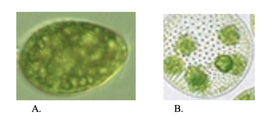
This supergroup includes a large group of algae, specifically the green algae from which higher plants have emerged. This supergroup consists of several major groups, such as: red algae that contain phycoerythrin (photosynthetic pigment), green algae with plant-type chloroplasts, and land plants (discussed in later labs). The microscopic green algae include unicellular as well as various colonial, coccoid, and filamentous forms of flagellates (usually with two flagella per cell), that all contain chloroplasts. Genera within the green algae include Euglena, Chlamydomonas, Volvox, Ulothrix, and Spirogyra.
There are about 6000 species of green algae; many species live most of their lives as single cells, while other species form colonies or long filaments. Algae employ simple reproductive structures and lack the extensive vascular structures characteristic of higher plants. Eukaryote organisms are capable of oxygenic photosynthesis. They are classified into different groups on the basis of morphology, types of chlorophylls, carbon reserve storage materials, cell wall composition, and habitat. Although many algae, like the higher plants, are non-motile, they may have motile reproductive cells.
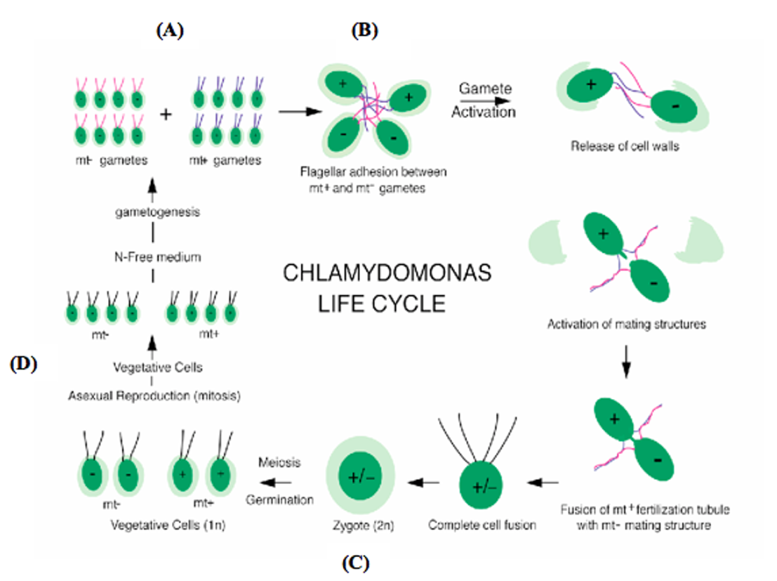
This organism usually exists in a haploid stage, having just one copy of each chromosome (like a mammalian gamete). When mating types plus (mt+) and minus (mt-) meet (Figure 6A), flagellar adhesion and gamete activation are initiated (Figure 6B). They join their cytoplasm (plasmogamy) and their nuclei fuse (karyogamy) to form a diploid zygote (Figure 6C). The zygote is the only diploid cell stage in the life cycle, and it eventually undergoes meiosis to form a tetrad, germinating to form four new Chlamydomonas cells, two mt+ and two mt- following the Mendelian genetics. (Figure 6D). When vegetative cells are grown in a nitrogen-free growth medium, Chlamydomonas cells undergo gametogenesis and develop into gametes (Figure 6A).
Activity 4: Archaeplastida Observations
Answer the following questions. (The following questions are from the sources attributed below.)
1. View the green algae Chlamydomonas reinhardtii specimens available and then answer the questions below.
a. What pigment does green algae use for photosynthesis?
b. Name and describe the characteristics of one green algae specimen.
2. View the red algae specimens available and then answer the questions below.
a. What pigment do red algae use for photosynthesis?
b. Name and describe the characteristics of one red algae specimen.
V. Supergroup Unikonta
This supergroup consists of several major groups, such as Amobozoans, with tube-shaped pseudopodia, and highly variable Opisthokonts. Some examples of the Amobozoans include Amoeba and Dictyostelium (slime molds). This group consists of amoeboid protozoa that use pseudopodia for locomotion. Examples include amoebae (Entamoeba histolytica is the cause of amoebiasis) that do not have an outer covering on the pseudopodia (naked) or have a protein or mineral coating over the pseudopodia. Examples of the Opisthokonts include choanoflagellates, animals, and fungi (discussed in future labs).
Activity 5: Unikonta Observations
View the tubulinid specimens available (Amoeba) and then answer the following questions. (The following questions are from the sources attributed below.)
1. What structure does Amoeba use to move?
2. Is the Amoeba single or multi-celled?
3. The Amoeba contains contractile vacuoles that help maintain water balance through osmosis. Can you locate any on your specimen?
VI. Post-lab Activities
For the post-lab activities, write your answers in your lab report.
Activity 6: Eukaryote Phylogeny
1. As you have learned, a group must be monophyletic in order to be considered a kingdom. Watch the video below from the beginning to 1:10. Based on the tree shown (0:23–0:42), do you think that protists are truly a kingdom? Explain how you know.
2. Watch the video below and then answer the questions that follow in your lab report as you go. Use the timestamps given to help you find the answers. The video is just under nine minutes long.
The video explains that each domain can be divided into subgroups (0:42). Ideally, the next level after the domain is kingdoms, but since these have not been worked out for many eukaryotes, biologists use “supergroups” while the work of classification is in progress.
1:24 The video uses the classification system with five supergroups of eukaryotes, each of which is assumed to form a clade. What does the word “clade” mean?
2:39 Do you agree with the definition “protists are unicellular eukaryotes”? Why or why not? (See also 3:23–3:50 in the video.)
4:55 Different biologists make different phylogenetic trees and different supergroups for eukaryotes. Why do you think this is so? (See also 5:11.)
5:43 What is the definition of polytomy? (Watch until 6:21.) Why must the eukaryote tree include polytomy?
6:37 Why is it easier to figure out the evolutionary relationships for the more specific subgroups towards the right in the diagram?
7:30 How can you differentiate among the various supergroups—in other words, how can you tell them apart, according to this video?
8:00 Do you think the method in the previous question will work well for classifying an unknown protist? Why or why not?
8:16 Why does the narrator say that the term algae is not useful? In what way?
Activity 7: Classifying Protists
This activity is based on some helpful online resources. Your instructor may have you watch these videos before class, or you may watch them together during lab.
1. Visit the Pondlife website and skim the page Microbes in Motion, which contains thirteen short videos, each featuring a different microbe. Twelve of these are common protists collected from ponds and marshes right here in New York City (the other is a bacterium). These videos should give you a good idea of what a variety of protists look like.
2. Watch the video below with particular attention to the section on protists (4:34–6:45) and then answer the questions that follow, using what you learned at the indicated timestamps in the video.
6:10 What would be the impact on the world if diatoms suddenly went extinct?
6:46: What is the difference between protists and microscopic animals? How can you tell them apart? Hint: it’s not size, and it’s not multicellularity. If you need another hint, go back to the video Kingdom of Protista above and watch 0:55–1:04 again.
3. Now that you have a better idea of what a protist is and how to differentiate it from microscopic animals, let’s test your expertise. Watch the video below and see if you can find at least two places where the filmmaker mistook an animal for a protist. Can you catch his mistakes? One caveat: this question does not apply the animals you see during the first minute of the video (bird, lizard, snake, fish, and humans). The animals that appear later on are tricky—they are microscopic, like many protists, which is why the filmmaker may not have known that they are animals.
Was identifying the animals in the video easy or difficult? What made it easy or difficult for you?
Activity 8: Summary Questions
Answer the following questions. (Some of the following questions are from the sources attributed below.)
1. How is the classification of protists different from that of other eukaryotes?
2. What does the current status of protist classification tell you about their evolution?
3. What can you conclude about diversity in the protists? (Is it greater or less than other eukaryotes, for example?)
4. What have you learned about the importance of protists in their ecosystems? What important roles do they play?
5. Which protists are most similar to green plants? Why?
6. You viewed several protista that exhibited movement. Give an example of a protist that used each of the following movement structures:
Flagella:
Cilia:
Pseudopods:
7. Give two examples of photosynthetic protists you viewed in the lab and state what pigment each uses for photosynthesis.
Attribution
Questions in this lab are adapted from “2.1: Microbiology and Protista Lab” by Lynette Hauser, licensed CC BY 4.0, and from Prokaryotes Lab (Biology 102) by Michael J. Gregory, Ph.D., licensed CC BY-NC-SA 4.0.
