Week 5: Fungi
Objectives
- Describe fungal classification into phyla, and provide a phylogeny of Kingdom Fungi.
- In a sentence or two, describe the characteristics of the three largest phyla in the Kingdom of Fungi (Zygomycota, Ascomycota, Basidiomycota).
- Using images, explain the life cycle of typical multicellular fungi.
- Give three examples of how humans benefit from specific uses of fungi.
I. Fungal Classification and Phylogeny
The study of fungi is termed mycology. Members of the Kingdom Fungi are eukaryotic organisms, which have characteristics that distinguish them from prokaryotes and from the rest of the eukaryotes. For example, fungi possess nuclei with a nuclear envelope and an endomembranous system that includes the endoplasmic reticulum and Golgi bodies. Fungi exhibit diverse morphological features that have been used in their classification, but all members do share some characteristics. For example, fungi have a cell wall composed of a durable polysaccharide called chitin (also found in arthropod exoskeletons). If not pathogenic, fungi can form symbiotic relationships or act as decomposers. Fungal species may be unicellular (e.g., yeast) or multicellular (e.g., mushrooms). Human fungal infections are called mycoses. Reproduction in fungi may include both sexual and asexual reproduction, although some species reproduce strictly asexually.
Before coming to the lab, your instructor may request that you review “Reading: Fungi.”
And, for more information, watch the following videos (optional):
Key Terms
| Term | Definition |
| Symbiosis | A type of ecological interaction in which two species are closely associated, usually temporally and spatially |
| Mutualism | A type of symbiosis in which both species involved in the interaction benefit |
| Commensalism | A type of symbiosis in which one organism benefits while there is no apparent benefit or disadvantage to the other |
| Parasitism | A type of symbiosis in which a pathogen benefits from a host (who is harmed) |
| Pathogen | Parasites that can cause disease |
| Hyphae | Fungal structures that serve as building blocks in multicellular fungi. Hyphae may be organized into feeding structures called mycelia, or may form a reproductive structure called a fruiting body. |
| Plasmogamy | The union between two cell membranes without the union of nuclei |
| Heterokaryotic stage | A stage in fungal reproduction that occurs between plasmogamy and karyogamy |
| Karyogamy | The union of nuclei in heterokaryotic cells, following plasmogamy and the heterokaryotic stage |
Classification/Phylogeny of Fungi
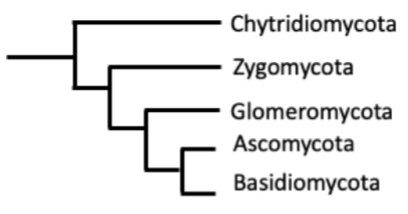
The phylogenetic relationships between the fungal classes are still under investigation. With the sequencing of complete rRNA of many different species, the classification is ever-changing and new phylogenetic trees have been generated based on this data (Tedersoo et al., 2018). The general consensus among phylogeneticists that study Kingdom Fungi is that they form a monophyletic group. The ancestor of the fungi was likely an aquatic organism that resembled a flagellated single-celled protist belonging to the group we currently know as Opisthokonta (Unikonta). Figure 1 summarizes the phylogeny of Kingdom Fungi.
Here are some highlights from each group.
| Phylum | Highlight |
| Chytridiomycota | The most primitive of the fungi, with approximately 1000 species. They are known for their flagellated spores. |
| Zygomycota | They form a freeze-resistant reproductive structure called a zygosporangium. This group also has about 1000 species. |
| Glomeromycota | They are known for forming arbuscular mycorrhizae (symbiotic relationships with plant roots). This is the smallest group at only about 160 species strong. |
| Ascomycota | Known as sac fungi, form asci (singular ascus, a sac-like structure), which contain haploid ascospores. This is the biggest group containing some 65,000 species. |
| Basidiomycota | Also known as club fungi due to their club-shaped reproductive structures (basidia) containing haploid basidiospores. About 30,000 species strong. Some species are edible. |
Life Cycle of Fungi
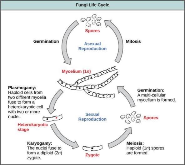
All unicellular fungi (such as yeast) reproduce by asexual, mitotic cell division. Multicellular fungi can reproduce via a combination of sexual and asexual reproduction (Figure 2). The sexual portion of the life cycle begins with two different parent mycelia, which grow from spores. A mycelium is a collection of structures called hyphae (long, branching filaments that increase surface area). In fungi, there are no males and females—only genetically distinct mating types (often designated + and -). Two cells from the different mating types join together, but their nuclei remain separate, each still enclosed by its nuclear envelope. This fusion of two parent cells is called plasmogamy and results in a cell with two genetically different nuclei. Such cells are called heterokaryotic and may continue to divide and produce daughter cells that also contain two nuclei each. Some fungi remain in this heterokaryotic stage for a long time, but eventually, the nuclei combine to form diploid cells with a set of chromosomes from each parent mycelium. This fusion of the heterokaryotic nuclei into diploid nuclei is called karyogamy and produces a zygote. The zygote undergoes meiosis shortly after karyogamy, forming haploid spores which can then disperse and germinate into new mycelia to continue the life cycle. The asexual part of the life cycle results in the production of haploid spores by mitosis (Figure 2). Approximately 20,000 species of fungi, collectively known as the Deuteromycota, reproduce strictly asexually. Deuteromycota contains species from both Ascomycota and Basidiomycota.
Before lab, watch the following video to help visualize budding:
II. Fungal Diversity
Activity 1: Zygomycota
Introduction to Zygomycota
Before coming to the lab, watch the following video on Zygomycota:
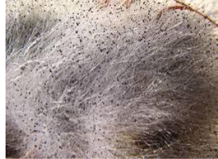
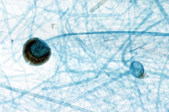
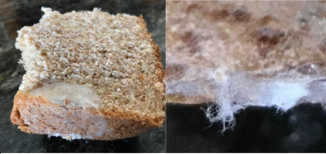
Rhizopus is a member of the Zygomycota. They are mainly saprotrophic, meaning that they feed on dead organisms, and therefore play a major role in decomposition. Look at the images of Rhizopus below.
Exploratory observation of Zygomycota
Visual observation of bread mold
Go to the display table to observe the petri dish with Rhizopus stolonifer (bread mold). There are three Petri dishes, so your group may have to wait before observing the petri dish with the specimen on it. If the petri dish is in use, please go to the next exercise (Zygomycota microscopy) and return to this exercise when a petri dish becomes available. Note: Do not open the petri dish used in the demo.
Answer the following questions.
1. Based on what you learned and observed, what do you think the black granulation (dots) represents?
2. Go to the display table and locate common Rhizopus species. What does it grow on?
Zygomycota microscopy
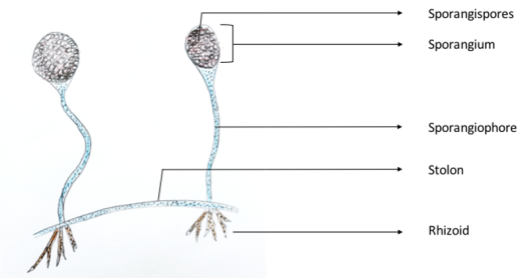
Go to the display station and retrieve the slide for observation of Rhizopus sporangia (bread mold). Compare and identify what you observe under the microscope to the drawing in Figure 5, and then answer the questions and complete the activities below.
1. What are the functions of each structure in Figure 5?
Sporangium:
Stolon:
Rhizoid:
2. Draw a picture of the zygospores as you viewed them under the microscope.
3. Zygomycota (bread mold): View the prepared slides and the culture dish of the zygospores and sporangia. (The following questions are from the source attributed below.)
a. What kind of reproduction is used by the Zygomycota?
b. Is the zygospore diploid or haploid?
Activity 2: Ascomycota
Introduction to Ascomycota
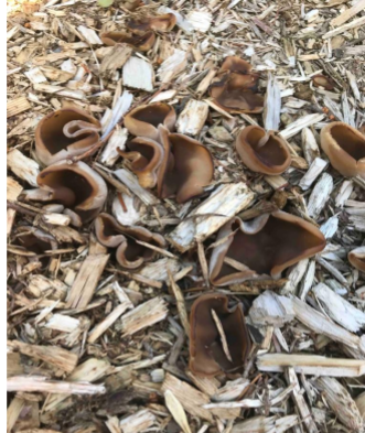
Ascomycota belongs to the largest phylum of Fungi with over 64,000 species. Together with the Basidiomycota, they form the subkingdom Dikarya. Ascomycota is also known as sac fungi.
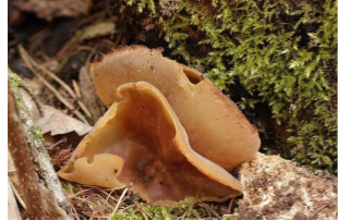
Examine the images of Peziza and other ascomycetes below.
Exploratory observation of Ascomycota (sac fungi)
Visual observation of Eurotium chevalieri
Go to the display table to observe the petri dish with Eurotium chevalieri (bread mold). There are three Petri dishes, so your group may have to wait before observing the petri dish with the specimen on it. If the petri dish is in use, please go to the next exercise (Visual and microscopic observation of Pezzia) and return to this exercise when a petri dish becomes available. Note: Do not open the petri dish used in the demo.
Describe your exploratory observations.
Visual and microscopic observation of Pezzia (cup fungi)
Go to the display table to observe the petri dish with Pezzia (cup fungi). There are three Petri dishes and slides.
Answer the following questions. (Some of the following questions are from the source attributed below.)
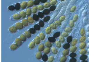
1. What are the small, oval, green/black structures called in Figure 8? (Consult the caption for help.)
2. What is the name of the structure that contains the small, oval, green/black structures?
3. View the slides available on Aspergillus or Peziza. Can you find any conidiospores?
4. Are conidiospores used in sexual or asexual reproduction?
5. Draw a picture of the conidiospores as you viewed them under the microscope.
Some of the fungi that are most important to humans include common yeast, Saccharomyces cerevisiae, which is used in the production of bread and beer and is classified in Phylum Ascomycota. (The following questions are from the source attributed below.)
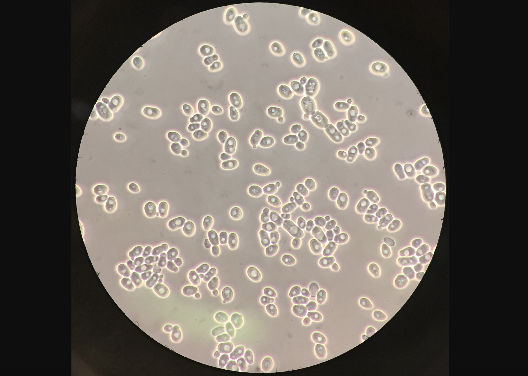
6. Observe figure 10 and refer to the fungi life cycle diagram to answer the questions below.
a. Are yeast single or multi-celled?
b. Does yeast reproduce asexually or sexually?
c. Are you able to view budding, the asexual reproductive process of yeast?
d. When yeast reproduces sexually, what is the name of the diploid cell that is formed?
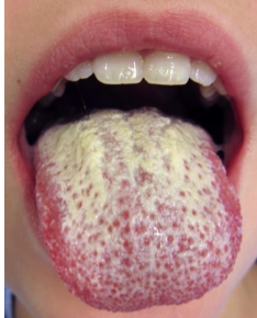
Some Ascomycota species are opportunistic pathogens of humans. For example, candidiasis (thrush), is caused by the fungus Candida albicans.
In your free time, watch the following interesting video on Candidiasis infections and treatment.
Figure 11 shows one example of how thrush may look.
Activity 3: Basidiomycota
Introduction to Basidiomycota
Before coming to the lab, watch and take notes on the following videos:
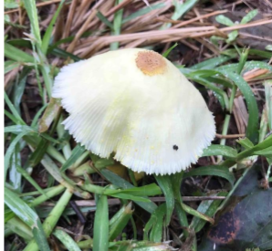
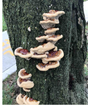
Basidiomycota is the second largest class of fungi. Collectively they are referred to as the club fungi. Proceed to the display table and observe the Basidiomycota fruiting body and examples in the images below.


Many basidiomycetes are useful to humans as food, but fungi from this phylum and the other phyla have many other uses that are beneficial to humans. Think of an example of a product in your house that could not be made without a fungus (don’t use mushrooms or other food—find something else!). It’s OK if the fungus you choose is not from Phylum Basidiomycota. How are fungi involved in the item’s production?
Exploratory observations of Basidiomycota (club fungi)
Observation of common club fungi (portabella and Coprinus)
Answer the following questions. (The following questions are from the source attributed below.)
1. View the portabella mushroom specimens available in the lab. Do not dissect them. See if you can find the gills on the underside of the basidiocarp.
a. Name the specific spores formed by the mushroom in the gills.
2. View Figure 17, the cross-section of the Coprinus mushroom.
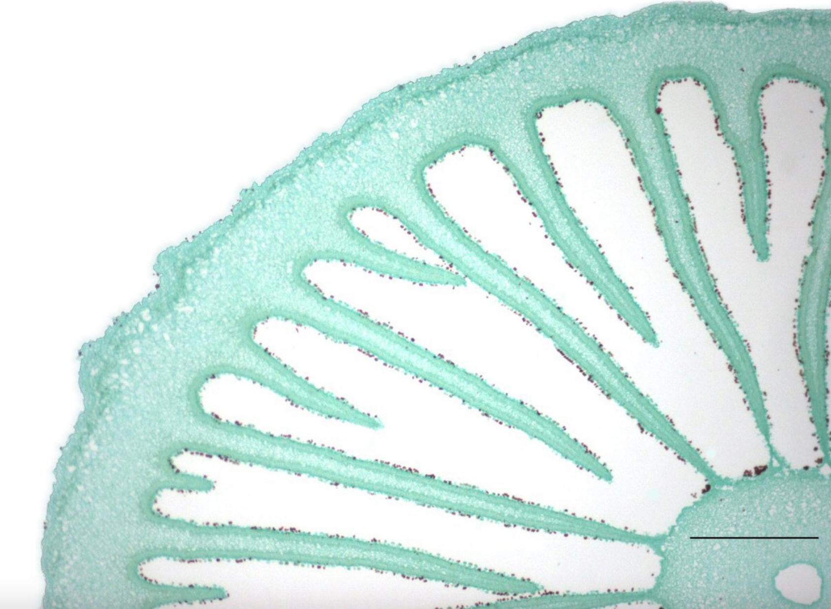
a. Can you locate the basidiospores?
b. Name the specific stalk that the basidiospores attach to.
Activity 4: Imperfect Fungi (Deuteromycota)
Deuteromycota is composed of approximately 20,000 species belonging to the Ascomycota and the Basidiomycota.
Exploratory observation of imperfect fungi (Deuteromycota)
Observation of Penicillium notatum
Proceed to the demonstration table to observe the demo and slides of Penicillium.
What is the significance of Penicillium?
View the Penicillium slides and answer the questions. (The following questions are from the source attributed below.)
1. Name the specialized stalks that the asexual spores attach to.
2. Draw a picture of the Penicillium specimen as you viewed it under the microscope.
Activity 5: Mutualistic Fungi
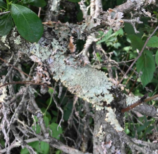
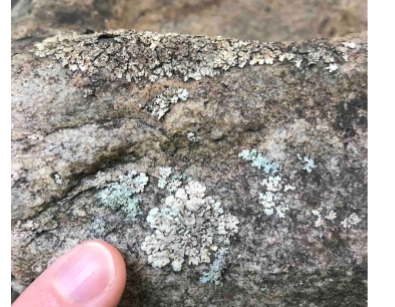
Fungi can form mutualistic relationships with unicellular photosynthetic organisms. These are generally called lichens.
Exploratory observation of mutualistic fungi (lichen/mycorrhizae and Rhizobium)
Observation of lichen
Proceed to the demonstration table to observe the lichens and view the figures below.

1. Name two types of photosynthetic organisms that can form mutualistic relationships with a fungus to form lichens:
2. What does the fungus bring to this mutualistic relationship?
3. What does the photosynthetic organism bring to this relationship?
There may or may not be live specimens of the lichens to view in the classroom. If live specimens are present, please look at them and answer these questions. (The following questions are from the source attributed below.)
4. What type of lichen has the algae dispersed throughout?
5. What type of lichen exhibits the fastest growth?
6. What type of lichen grows in a circular pattern forming lobes?
7. View the lichen thallus slide under the microscope. Use the space below to draw a picture of the lichen thallus as you viewed it under the microscope. On your picture try to label both the fungi and the algae.
III. Post-lab Questions
Answer the questions below to summarize the lab activity. (The following questions are from the source attributed below.)
1. What is the domain of fungi?
2. How do fungi obtain energy?
3. What is the reproductive structure of fungi? It’s not sperm and egg!
4. In the lab activity, which groups of fungi prefer to reproduce asexually? Which groups of fungi tend to exhibit sexual reproduction?
5. A lichen is a mutualistic relationship between what two organisms?
Attribution
Questions in this lab are adapted from “3.1: Fungi Lab” by Lynette Hauser, licensed CC BY 4.0.

.jpg)