Week 8: Animals I — Invertebrates
Objectives
- Distinguish members of the Kingdom Animalia from their closest living relatives (Choanoflagellates and Fungi).
- Explain the basic body plan of members in the Kingdom Animalia.
- Identify members of the Phyla Porifera, Cnidaria, Platyhelminthes, Rotifera, Annelida, Mollusca, Nematoda, Arthropoda, and Echinodermata.
- Compare two types of invertebrate life cycles.
- Compare the structure and function of invertebrates.
This lab begins with an introduction to Metazoans—the animal kingdom—including their common characteristics and taxonomy. It then includes nine activities, each focused on one of the major phyla of invertebrates. Activities each include a pre-lab introduction and one or more in-lab exercises.
- I. An Introduction to Metazoans
- II. Invertebrates
- III. Superphylum Lophotrochozoa
- IV. Superphylum Ecdysozoa
- V. Superphylum Deuterostomia
- VI. Post-lab Questions
This lab includes the following diagrams to be printed out and labeled.
I. An Introduction to Metazoans
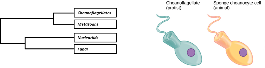
Metazoans (animals) evolved over 600 million years ago. Their closest living relative based on molecular and morphological evidence is the choanoflagellates (flagellated protist, Figure 1). The best hypothesis to support the split from their common ancestor is the development of an extracellular matrix and communication junctions. These two features allowed the first metazoans to form cell colonies, which would eventually develop true multicellularity and specific body plans and tissues specialized for movement, nutrition, reproduction, and body support.
Key Terms
| Term | Definition |
| Blastocysts | A multicellular structure that forms during the early stage of embryonic development. The cells of this structure will begin to form an inner cell mass that becomes the embryo. |
| Gastrulation | A period during embryonic development when the germ layers (endoderm, mesoderm, ectoderm) are formed. |
| Coelom | A term used to describe a triploblastic body cavity that develops from the mesoderm. |
| Acoelom | A term used to describe triploblasts without a body cavity. |
| Pseudocoelom | A term that means “false cavity.” This triploblastic cavity is developing from mesoderm and endoderm. |
| Mesohyl | The extracellular matrix of the sponge that houses several cell types. |
| Diploblast | Organisms that are derived from two embryonic germ layers (ectoderm and endoderm). |
| Triploblast | Organisms that are derived from three embryonic germ layers (ectoderm, mesoderm, and endoderm). |
| Hermaphrodite | An organism with a complete or partial set of male and female sex organs or gametes (ova or sperm). Common in simple invertebrates. |
| Cephalization | Is a word associated with the formation of a head. It can also imply the development of a mouth and nervous tissue. |
| Exoskeleton | Outer skeleton is found in arthropods. |
Classification
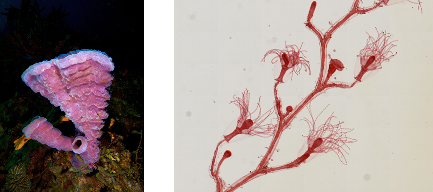
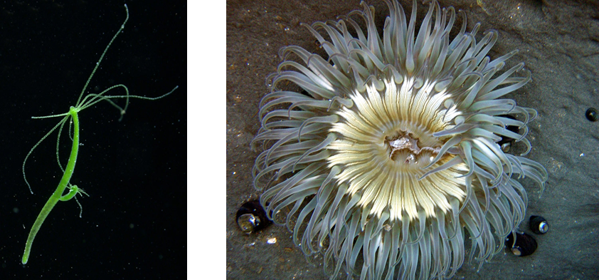
By looking at a more detailed phylogenetic tree (Figure 2), it can be said the Metazoan clade is very diverse. Some group members have similar morphological features, while simple animals, such as the sponges, Obelia, Hydra, and corals (Figure 3), look similar to plants. Based on morphology, you might begin asking yourself—What separates these simple animals from other eukaryotes you have previously discussed, like plants and fungi? It’s simple! Members of this clade lack cell walls.

All animals share similar characteristics that separate them from their closest relative, the choanoflagellates; see Table 1.
| Characteristic | Description |
| Multicellularity | A condition in which an organism is composed of cells that carry out multiple, coordinated functions. In animals, these cells exist in an extracellular matrix composed of collagen fibers. |
| Motility | The ability to move in response to a change in the environment (search for food, respond to a change in temperature, and mating) or disperse (larva of sessile animals). |
| Heterotrophic | Animals cannot synthesize most organic molecules. Therefore, they have to feed off of the living and/or non-living organic material. |
| Sexual reproduction | Most members of the animal kingdom are diploid and reproduce sexually. This means that most of their life cycle will exist in the diploid stage, while only the gametes (sperm and egg) are haploid. However, there are exceptions to this:
|
Pre-lab Activity
Before lab, watch the videos below to see how animals are further classified. Afterward, use the information from the videos to complete a chart like the one below. If a feature is not a characteristic of a particular phylum, either leave it blank or place a line through it.
Note: This video also discusses the classification of vertebrates, next week’s subject.
| Phylum | Motility (sessile or motile) |
Type of symmetry (asymmetry, radial symmetry, bilateral symmetry) |
Presence of tissues (none, diploblastic, triploblastic) |
Body cavity (acoelom, pseudocoel, coelom) |
Origin of the mouth (Protostome or Deuterostome) |
| Porifera | |||||
| Cnidarian | |||||
| Platyhelminthes | |||||
| Rotifera | |||||
| Annelida | |||||
| Mollusca | |||||
| Nematoda | |||||
| Arthropoda | |||||
| Echinodermata |
II. Invertebrates
Activity 1: Phylum Porifera
Simplest of all animals, the sponge has an asymmetric morphology composed of specialized cells that work together. Though the previous statement is also the biological definition of tissue, the sponge lacks “true” tissue.
Before lab, read the overview of Porifera linked below and then watch the video that follows.
Pre-lab: Basic anatomy of the sponge
Answer the following questions.
1. Why do you think animals in the Phylum Porifera lack “true” tissue?
2. Fill in a chart like the one below on sponge morphology.
| Name of cell | Location | Description/Morphology | Function |
| Choanocytes | |||
| Amoebocytes | |||
| Pinacocytes (epidermal cells) | |||
| Collagenocytes | |||
| Porocytes | |||
| Sclerocytes |
3. Download and print out a copy of Figure 4 below, and then label the diagram using the following terms: Osculum, Ostia, Spongocoel, Mesohyl. Using arrows, draw the direction in which water flows into and out of the sponge.
4. Remember, these are simple organisms; therefore, they lack complex organ systems (digestive, circulatory and respiratory, etc.). How would a sponge carry out gas exchange, circulation, and excretion?
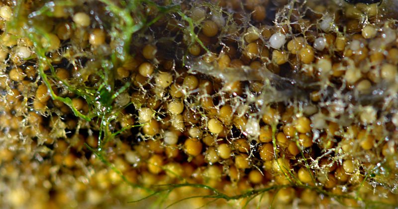
5. Sponges can reproduce sexually and asexually. Freshwater sponges do not use the typical means of asexual reproduction such as fragmentation or budding. However, they produce gemmules (Figure 5).
a. What are gemmules?
b. Why would a sponge produce gemmules?
In the lab: Observation of commercial sponges (preserved)
On your lab bench, locate the invertebrate box that contains various preserved specimens of invertebrates. Locate the preserved commercial sponge(s). And then answer the following questions (adapted from the source attributed below).
1. Dry sponge specimens of various species are located in your invertebrate box. If your box has one specimen, you may need to observe specimens from other groups. Please make observations on the available specimens and fill in a chart like the one below.
| Name of specimen | Physical description | Sponge structures visible (osculum, other pores, spicules) |
2. What type of symmetry is displayed in the sponge specimens?
Activity 2: Phylum Cnidaria
Before lab, read the overview of Phylum Cnidaria (all four sections).
All cnidarians have specialized cells called cnidocytes that are located in their tentacles and around their mouth. Cnidocytes contain specialized organelles called nematocysts.
Also watch this video on how jellyfish sting.
Pre-lab: Basic anatomy of the sponge
Answer the following questions.
Morphology
Two distinct body plans exist: medusa and polyp.
1. Use information from the reading to draw these three cnidarians in your lab notebook: jellyfish, hydra, and obelia. Using a table like the one below, identify the body plan (medusa, polyp, or both) and movement (motile or sessile).
| Jellyfish | Hydra | Obelia | |
| Body plan | |||
| Movement |
2. Think about how the hydra feeds. What is the benefit of having radial symmetry?
3. What is the function of the nematocyst?
4. Does the hydra have a nervous system? Explain.
Reproduction
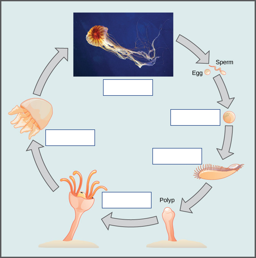
5. Download and print out a copy of Figure 6 below, and then label the diagram. Place your answers in the boxes.
In the lab: Observation of hydra feeding
In this activity, you will observe Hydra feeding of brine shrimp larvae. You will need the following items to prepare the slides for your observation:
- Glass slides
- Cover slip
- Dropper pipet
- Methylcellulose
The specimens are found at the demo station.
To prepare a wet mount
- Obtain a clean slide.
- Use a clean dropper pipet to gently remove one hydra from the sides of the container.
- Place a drop of solution with your hydra onto your slide.
- Add a small drop of methylcellulose on to the slide with the hydra. Methylcellulose is used to slow down the brine shrimp for observation.
- Use a new dropper pipet, place a small drop of the sample containing the brine shrimp onto the slide.
- Observe your newly prepared wet mount under the dissecting microscope and record your findings in the lab notebook.
Answer the following questions (adapted from the source attributed below).
1. Does the hydra illustrate the polyp or the medusa stage?
2. How many germ layers does the hydra contain?
3. Can you find the hydra tentacles? How many tentacles does your hydra specimen contain?
4. Name the stinging cells present on the tentacles that are unique to cnidarians.
5. Describe the movement of the hydra if live specimens are available.
6. Before lab, you watched the video that shows a jellyfish discharging the nematocyst cells: Nematocysts Firing. What did you notice that surprised or interested you?
III. Superphylum Lophotrochozoa
Activity 3: Phylum Platyhelminthes
Members of this phylum exist as either free-living or parasitic entities. Two platyhelminths we will observe are Planaria and tapeworms. Before lab, review “Reading: Flatworms.”
Pre-lab: Background on free-living and parasitic platyhelminth
Planaria: Free-living platyhelminth
Watch the video below, and then answer the questions that follow.
1. Does the planarian show signs of cephalization? Explain.
2. Is the digestive system complete or incomplete?
3. If you expose planarians to light, how do they react, and why?
4. What is the function of the flame cells?
5. What are zooids, and how do they form?
Tapeworm and river fluke: Parasitic platyhelminth
Read about the tapeworm’s life cycle on the CDC website and watch the video below about T. saginata. You do not need to listen to the diagnosis part of the video. And then answer the questions that follow. (Questions 6, 9, 11, and 12 are adapted from the source attributed below).
6. Name two livestock that can be infected by tapeworms.
7. Since cattle and pigs are not purposefully eating feces, how would the proglottid find its way into their systems?
8. How are humans infected by T. saginata or T. solium?
9. If a human is infected, where does the tapeworm live?
10. What is a proglottid?
11. Are tapeworms hermaphrodites?
12. What is a scolex?
13. What structures are located on the scolex to help the tapeworm attach to the host?
Liver flukes are an example of a parasitic flatworm. Read about the liver fluke’s life cycle on the CDC website and then answer the following questions (adapted from the source attributed below).
14. Where does the adult liver fluke live?
15. When the liver fluke egg hatches, what organism does it infect first?
16. Can humans become infected with liver flukes?
In the lab: Microscopic observation of planarians
In this exercise, you will be observing live and preserved planarian flatworms. Based on your observations, answer the following questions (adapted from the source attributed below).
1. Observe a live planarian flatworm, if available, under the dissecting microscope. Transfer one planarian from the culture container to a slide with a plastic pipette, and add a bit of water from the culture container. You will return the specimen to the culture container when finished observing it.
a. What type of symmetry does the planarian display?
b. Does the planarian exhibit cephalization?
c. Can you locate the planarian eyespots? What do the eyespots sense?
2. Examine the prepared slides of planarians with a compound light microscope. Make sure you can identify the pharynx, the eyespots, and the flame cells.
a. Does the planaria have a complete or incomplete digestive system?
b. What is the function of the flame cell?
c. Are planaria hermaphrodites?
Activity 4: Phylum Rotifera
Before lab, watch the video below of rotifers under the microscope. Rotifers are different from the previous phyla because they are sexually dimorphic—females are always larger than the males.
In the lab: Microscopic observation of the Rotifer
In this activity, you will observe the Rotifer. You will need the following items to prepare the slides for your observation:
- Glass slides
- Cover slip
- Dropper pipet
The specimens are found at the demo station.
To prepare a wet mount
- Obtain a clean slide.
- Use a clean dropper pipet to take a sample from the rotifer culture container.
- Place a drop of solution with the rotifer culture onto the slide and place a cover slip onto the slide.
- Observe your newly prepared wet mount under the compound light microscope and record your findings in the lab notebook.
Answer the following questions based on your observations.
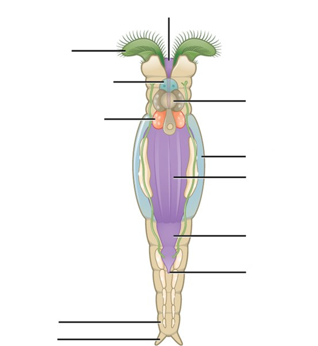
1. Download and print out a copy of Figure 7 below, and then label the diagram. Your instructor may also have you draw what you observe. Be sure to label your drawings with the magnification used and an approximate scale, as well as the name of the species.
2. Name two modes of motility in rotifers.
3. What is the mastax?
4. Describe the function of the corona.
Activity 5: Phylum Annelida
Before the lab, familiarize yourself with the Phylum Annelida by reading and taking notes on “Reading: Annelids.”
Pre-lab: Anatomy of segmented worms
Use your notes from the reading on Annelids to answer the following questions.
1. Define the following terms.
Hydrostatic skeleton –
Setae –
Clitellum –
Gizzard –
2. What is the evolutionary significance of segmentation?
3. Describe how earthworms can burrow and move on land without having legs.
In the lab: Earthworm dissection
In this activity, you will dissect the earthworm. You will need the following items to dissect the earthworm:
- Scissors – sharp dissecting scissors
- Blunt probe
- Dissecting pins (Do not discard.)
To dissect the earthworm
- Place the earthworm on the dissecting tray.
- Observe the external anatomy of the earthworm. Locate the mouth, setae, dorsal vein, and the clitellum.
- Using the dorsal vein at a marker for dissection by making sure it is facing you.
- To stabilize the specimen, use two dissecting pins and place one on each end.
- Using the dissecting scissors, make a shallow incision midway on the clitellum.
- Once you have made the cut, gently begin cutting and pinning the skin as you cut towards the anterior and posterior end. Make sure you do not cut through the dorsal vein.
- Use the model on display to assist with identifying the internal organs.
Answer the following questions (adapted from the source attributed below).
1. What type of symmetry does the earthworm display?
2. Does the earthworm exhibit cephalization? How can you tell?
3. Does the earthworm exhibit segmentation? Is it internal, external, or both?
4. Examine the live leeches on display, if available. Do not remove them from the container, and do not put your fingers into the container. Is segmentation visible? In what ways do the leeches differ from the earthworms?
Activity 6: Phylum Mollusca
Before the lab, watch the video below on the classification of mollusks.
Pre-lab: Overview of the phylum mollusca
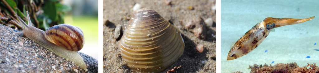
1. Mollusks are classified into three main classes: gastropods, bivalves (also called pelecypods in the video), and cephalopods. In a table like the one below, explain the meaning of the names and describe how each one moves.
| Class | Meaning | Locomotion (motility) |
| Gastropoda | ||
| Bivalvia (Pelecypoda) | ||
| Cephalopoda |
2. Looking at the picture below (Figure 8), it is safe to say that not all members of this phylum show signs of cephalization. Why do animals in the Class Bivalvia (Pelecypoda) lack cephalization?
In the lab: Dissection of the Bivalvia.
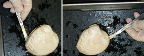
Before the lab, watch the two videos below on clam dissection.
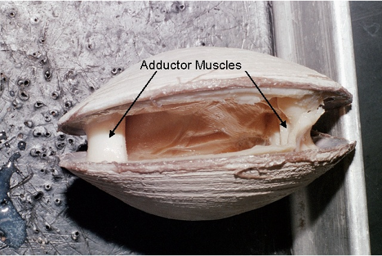
The image below (Figure 9) may also help. Identify the gills, adductor muscles, foot, labial palps, and mantle. What is the function of each structure? How is the shape or form adapted to that function? Complete a table like the one that follows.
External anatomy
- Obtain your bivalve—clam. You will notice a hump on each valve which is called the umbo. The umbo is the oldest part of the clam.
- Moving away from the umbo, you will notice the lines or ridges (growth ridge). As the clam grows, it will deposit a composite of proteins and calcium salts yearly. Like a tree, you can use the growth ridges to determine the age of your clam by counting the darkest ring. Approximately, how old is your clam? __________
Internal anatomy
- Notice that the hinge is located between the two umbos. The hinge is at the back of the clam.
- Hold the clam with the umbo facing away from you.
- You will also notice a small piece of wood wedged between the two shells. This is here to assist you with your dissection.
- Take a look between the opening created by the wooden wedge. You will notice two very strong adductor muscles—anterior and posterior adductor muscles.
- Using your scalpel cut, cutting away from your hand, you will cut the first adductor muscle.
- Once both adductor muscles have been cut, you want to pull both shells apart to reveal the internal structures.
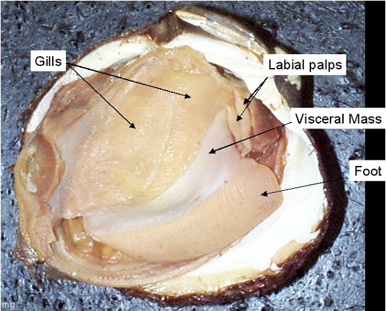
- Referring to Figure 9, try to identify the following internal structures:
- Gills
- Anterior foot retractor muscle
- Mantle
- Foot
- Labial palps
- Siphons
| Organ | Function |
| Gills | |
| Adductor muscles | |
| Foot | |
| Labial palps | |
| Mantle | |
| Siphons |
In the Lab: Dissection of the Cephalopoda
Dissect a squid following the procedure below. Make sketches in your lab notebook or lab report as directed by your instructor, and answer the following questions. (Instructions and directions are from the source attributed below). Find helpful diagrams and a step-by-step slideshow at Biology 11—Squid Dissection.
External anatomy
- Place the squid in the dissection pan with the mantle (major body part) facing away from you and the tentacles and arms towards you.
- Turn the squid so that the siphon faces you. It is located between the eyes. By expelling water through the siphon the squid can effectively move through the water.
- Notice the chromatophores on the mantle. They allow the squid to change color and blend into the environment.
- At the pointed tip of the mantle there are two fins that help stabilize and propel the squid
- Notice the eyes on either side-they are well developed and allow the squid to have excellent vision. You may wish to dissect out one of the eyes and open it with a scalpel to discover the lens.
- Distinguish between the tentacles and the arms. The tentacles are longer and are used to pass food to the arms.
- Count the number of arms. How many are there? ___________
- Notice the suction cups on both the tentacles and the arms. How does the distribution of the suction cups differ between these two structures?
- Pull back the arms and locate the beak or mouth in the middle. Use scissors to dissect out the beak, after you have examined the rest of the internal anatomy as instructed below.
Internal anatomy
- Use a pair of scissors and starting at the bottom of the mantle above the siphon cut one long incision up to the tip of the mantle. Be careful to lift up with the scissors while cutting to avoid cutting the internal organs.
- Spread the mantle open and try to identify the following internal structures:
- Feathery gills
- Heart, located at the base of each gill. Squid actually have three hearts!
- Gonads (ovary or testis), probably yellowish in color and long in shape running down the middle
- Cecum, a long, oval-shaped organ adjacent to the gonad. Part of the digestive system
- Ink sac, which looks like a small silver fish. If you find it, cut it out at both ends and you can extract some of the ink and try to write with it!
- The pen, which is all that remains of the shell. To try and find the pen, lift the head of the squid and place it down over the organs. You should notice a pointy area along the midline of the body, the tip of the pen. If you grasp the tip and pull the pen will release from the mantle. It resembles a transparent feather.
If required by your instructor, complete a chart like the one below based on your observations. In your answer, indicate how the shape or form of each organ is adapted to its function.
| Organ | Function |
| Beak | |
| Siphon | |
| Buccal mass | |
| Pen | |
| Brachial heart | |
| Systemic heart | |
| Nidamental gland |
IV. Superphylum Ecdysozoa
There are two phyla that we will discuss in this superphylum, the Arthropoda and the Nematoda. Read the overview of Superphylum Ecdysozoa (all four sections), which will help you answer the following questions. In addition, videos will be provided in each section.
Activity 7: Phylum Nematoda
Before the lab, watch and take notes on the following videos about nematodes. Disclosure: The first video contains images of parasitic infections in humans. If you are sensitive to graphic visual imagery, do not watch this video past the 3:26-minute mark.
Pre-lab: Intro to Nematode anatomy
Use the notes from the videos on the phylum Nematoda to answer the following questions.
1. Define cuticle.
2. Define ecdysis.
3. Since nematodes do not possess an exoskeleton like their closest relative, the arthropod, why do they fall under the superphylum Ecdysozoa?
In the lab: Microscopic observation of nematodes (vinegar eels)
In this activity, you will observe the vinegar eel. You will need the following items to prepare the slides for your observation:
- Glass slides
- Cover slip
- Dropper pipet
*The specimens are found at the demo station.
To prepare a wet mount:
- Obtain a clean slide.
- Use a clean dropper pipet to take a sample from the vinegar eel culture container.
- Place a drop of solution with the vinegar eel culture onto the slide and place a cover slip onto the slide.
- Observe your newly prepared wet mount under the compound light microscope and record your findings in the lab notebook.
Answer the following questions (from the source attributed below).
1. We will not be dissecting the Ascaris, but you should view the preserved specimens available in the lab.
a. What type of symmetry is seen in the roundworm?
b. Does it exhibit cephalization?
c. Ascaris is a parasite that infects humans, living in the intestines. What structure protects the nematode from being digested?
2. View the Trichinella slide (or slides of other preserved nematodes available in the lab). Trichinella is also a parasite. It can infect humans as well as other mammals like pigs, bears, and rodents. If untreated, it can lead to death.
a. What mammal tissue does this roundworm infect?
b. Draw a picture of the Trichinella as viewed under the microscope.
3. If available, view the live vinegar eel specimens. If there are no live specimens available, check out the video below instead.
a. Describe the movement of the vinegar eels.
b. Do they have a complete or incomplete digestive system?
Activity 8: Phylum Arthropoda
Phylum Arthropoda is by far the largest and most diverse phylum of animals. For that reason, this taxon is subdivided in subphyla. Recall from your textbook that there are four extant (living) subphyla of arthropods:
- Chelicerata (spiders, ticks, scorpions, horseshoe crabs, etc)
- Myriapoda (centipedes and millipedes)
- Hexapoda (insects)
- Crustacea (lobsters, crabs, shrimp, etc).
There is also one extinct subphylum, Trilobitomorpha (trilobites).
Before the lab, watch and take notes on the following video on arthropods.
Pre-lab: Intro to Arthropods
Use the notes from the videos on the phylum Arthrodpoda to answer the following questions.
1. List the three main characteristics of the arthropod body plan.
2. What type of circulatory system is present in this phylum?
3. List three types of respiratory structures found in arthropods.
4. Why do arthropods molt?
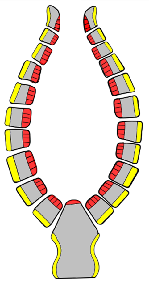
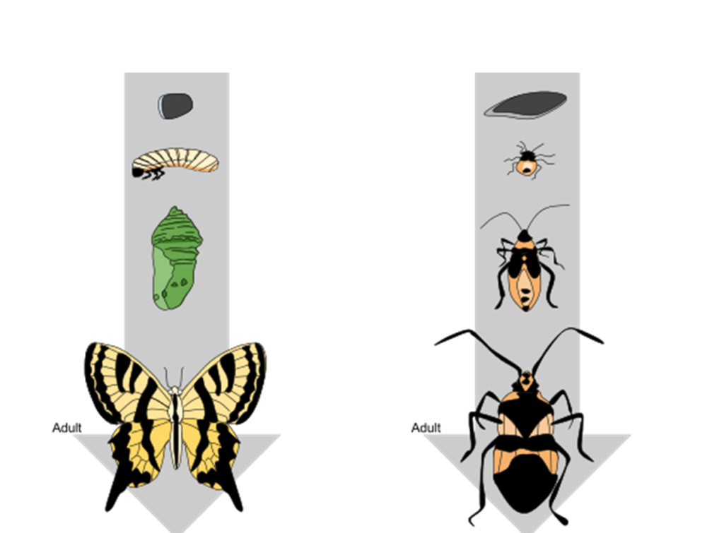
5. Download and print out a copy of Figure 10 below, and then label the diagram using the following terms:
-
- Complete metamorphosis
- Pupa
- Young nymph
- Larvae
- Incomplete metamorphosis
- Egg
- Later nymph
In the lab: Specimen observation of various preserved arthropods
On your lab bench, locate the invertebrate box that contains various preserved specimens of invertebrates. Locate the preserved species of arthropods, and then answer the following questions (adapted from the source attributed below).
1. View the preserved arthropod specimens available. Use a table like the one below to organize your observations.
| Name of specimen | Subphylum | Habitat (marine, freshwater, or terrestrial) | Number of jointed appendages | Number/type of body segments |
2. What type of symmetry is displayed in the arthropods?
3. Do the arthropods exhibit cephalization?
In the lab: Crayfish dissection
Before the lab, watch the two crayfish dissection videos below, the first focused on the external anatomy of a crayfish ad the second on its internal anatomy. As you watch, notice where the organs are located and, in a chart like the one below, jot down the function of each structure. Some functions may be found in your lab book or reading, as well.
Read the section “Crayfish Dissection” in “Reading: Arthropods” and see the guidance in Figure 11.
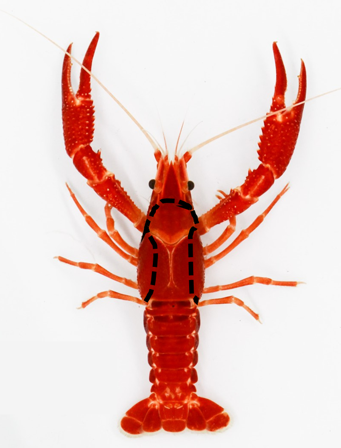
External anatomy
- Place the crayfish in the dissection pan. The first thing you will notice is the carapace – hard exoskeleton made of chitin.
- Turn the crayfish so that the legs are facing the tray. This is the dorsal view.
- The body of the crayfish is divided into the cephalothorax (fused head and thorax), abdomen, uropod and telson.
- In the cephalothorax region, locate the following structures:
- rostrum
- compound eyes
- antennae
- antennules
- Now, turn the crayfish over so that you have a better view of the legs. This is the ventral view.
- In the head region, behind the antennae, locate the mouth. You will notice that the mouth consists of multiple parts – mandibles (true jaw), maxilla and maxilliped.
- On the cephalothoracic region just below the mouth, notice the large appendage called the chelipeds or claws and 4 pairs of walking legs.
- On the ventral aspect of the abdomen, there are also 5 pairs of swimmerets. Crayfish exhibit sexual dimorphism or morphological differences between male and female. In the male crayfish, the first two swimmerets are enlarged for copulation, there is a sperm duct opening on the base segment of the fourth walking leg and the abdomen is narrow. Female crayfish have seminal receptacles between the fourth and fifth walking legs, at the third walking leg you will find the opening for the oviduct, and the tail is broader (wider) than the males.
Internal Anatomy
- Place the crayfish dorsal side up. You will notice that the lateral side (flaps) of the carapace near the legs are not as stiff. Using your scissors, carefully cut away the carapace to reveal the internal structures. See the dotted line in Figure 11 to help with cutting the carapace of the cephalothorax..
- Once the cut has been made, gently and slowly remove the carapace. You might need to use the forceps to aid in the removal of the recently cut carapace so as to not destroy the structure.
- After removing the carapace try to identify the following internal structures:
- Gills
- Brain
- Green gland
- Mandibular muscle
- Pyloric stomach
- Intestines
- Gastric mill
- Cardiac stomach
- Digestive gland
- Extensor muscle
Answer the following questions based on your observations. (Questions 2–8 are adapted from the source attributed below.)
1. Complete a crayfish anatomy chart like the one below. In your answer, indicate how the particular organ size and shape helps the organ fulfill its function.
| Organ | Function |
| Carapace | |
| Walking Legs | |
| Swimmerets | |
| Maxillipeds | |
| Uropod | |
| Gastric Mill | |
| Antennule | |
| Antenna | |
| Maxilla |
2. Do you have a male or female crayfish? How can you tell?
3. How many swimmerets does your crayfish have? Is that the same for all crayfish?
4. How many rows of gills does the crayfish have? (Lift up the top layer or row to see if there are more below.)
5. Where do the gills attach?
6. Can you find the stomach and the digestive glands? Where is each located?
7. What does the stomach attach to directly?
8. Try to locate the green glands. What is the function of this structure?
V. Superphylum Deuterostomia
Activity 9: Phylum Echinodermata
This last phylum will serve as a link between the phyla studied above and the phylum we will study next week, the Chordata. Echinoderms are more closely related to chordates than any previous phylum because Chordata and Echinodermata are both deuterostomes.
To familiarize yourself with members of this group before lab, review and take notes on the overview of Superphylum Deuterostomia (all three sections) and the following video.
Pre-lab: Introduction to Echinoderms
Use your notes from the video on the phylum Echinodermata to answer the following questions.
1. Name five Echinoderm classes. (This can be found in the video)
2. What type of symmetry is present in the larvae? And in the adult?
3. What is a water vascular system?
4. Let’s revisit the video on the differentiation between protostomes and deuterostomes. Now compare it to Figure 12 below.
a. What similarities do you see between what was described in the video and the gastrulas that are shown in Figure 12?
b. What type of symmetry is present during the larval stage?

In the lab: Specimen observation of preserved starfish
On your lab bench, locate the invertebrate box that contains various preserved specimens of invertebrates. Locate the preserved species of starfish.
Several preserved echinoderm specimens will be on display in the lab. Please make observations on the available specimens and fill in a table like the one below.
| Common name | Echinoderm class | Distinctive characteristics |
In the lab: Starfish dissection
Before the lab, read the step-by-step instructions and pictures found in Reading: Echinoderms.
After the dissection, answer the following questions (adapted from the source attributed below).
1. Remember the oral side (where the mouth is located) is actually on the underside of the starfish. The top surface is called the aboral side.
2. On the oral side make sure you find the mouth.
3. Also on the oral side in the center region of each leg look for the tube feet. Tube feet are used for locomotion powered by the water vascular system. How many rows of tube feet does your starfish have?
4. Try to differentiate between the spines and the skin gills. The spines are longer and are used for protection. The skin gills are smaller and used for gas exchange.
5. Find the sieve plate/madreporite on the aboral side. This is the water entrance point for the water vascular system used for movement.
6. The starfish has plates located underneath the skin for protection and support. What material comprises these plates?
7. The starfish has a two-part stomach, the upper pyloric stomach and the lower cardiac stomach. Can you differentiate between the two stomachs on your specimen?
8. In the starfish arms you should find both digestive glands and gonads. The digestive glands are brown and typically on top of the off white gonads. Make sure you can identify both structures.
VI. Post-lab Questions
To complete this lab and summarize what you have learned, please answer the following questions (adapted from the source attributed below).
1. Which phyla exhibit bilateral symmetry?
2. Which phyla have no true tissues?
3. Which phyla contain parasitic species?
4. Which phyla are coelomates?
5. Which phyla observed today are deuterostomes?
6. Which phyla exhibit cephalization?
7. Which phyla that you viewed today have specialized appendages?
8. Which phyla exhibit radial symmetry?
9. Which phyla are pseudocoelomates?
10. Which phyla have a complete digestive system?
11. Which phyla are multicellular?
12. Which phyla are asymmetrical?
13. Which phyla are acoelomates?
14. Which phyla have a shell?
15. Which phyla contain hermaphrodite organisms?
Attribution
Questions in this lab are adapted from “7.1 Invertebrate Lab I” by Lynette Hauser, licensed CC BY 4.0.

.jpg)
.jpeg)
.jpg)