Week 9: Animals II — Chordates
Objectives
- List characteristics found in the Subphylum Urochordata and Cephalochordata.
- List characteristics found in the Subphylum Vertebrata of Kingdom Animalia.
- List characteristics of each of the major tetrapod groups: amphibians, reptiles, birds, mammals; and provide examples of each.
- Identify homologous structures in vertebrates, and explain the functions of each structure.
- Identify representatives from the eight vertebrate clades, Agnatha, Chondrichthyes, Osteichthyes (comprised of Actinopterygii and Sarcopterygii), Amphibia, Reptilia, Aves, and Mammalia.
- Identify and list eleven organ systems in vertebrate animals, their main organs, and provide the major function(s) of each (integumentary, skeletal, muscular, nervous, endocrine, digestive, respiratory, cardiovascular, lymphatic/immune, urinary, & reproductive).
- Compare the life cycles of amphibians and mammals.
- Identify structures in dissected specimens of representative vertebrates (frog and fetal pig).
This lab begins with an overview of the invertebrate chordates and then continues with an overview of the vertebrates. The activities include the dissection of a leopard frog and investigation of its anatomy.
I. Invertebrate Chordates
Phylum Chordata includes the vertebrates, discussed below, as well as two subphyla of invertebrates: Urochordata and Cephalochordata. This section discussing these two subphyla is adapted from “Reading: Chordates” by Michael J. Gregory, Ph.D., licensed CC BY-NC-SA 4.0.
All animals in the Phylum Chordata have the following characteristics at some point in their life history:
- a dorsal, hollow nerve cord.
- a dorsal supporting rod called a notochord. This is replaced by a vertebral column in vertebrates.
- pharyngeal clefts (pouches). These develop into openings to the exterior (gill slits) in some chordates. Gill slits functioned as a mechanism for filter-feeding in primitive vertebrates. The gills of fish function in gas exchange.
- a postanal tail. In most other kinds of animals, the digestive tract extends the entire length of the animal.
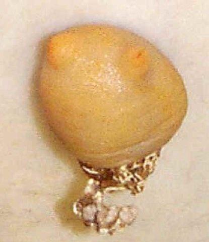
Subphylum Urochordata
Members of this subphylum are also called tunicates.
Larva
The larvae of tunicates resemble the ancestral chordate. The larval stage has chordate characteristics and looks like a tadpole. The free-swimming larva develops into a sessile, filter-feeding adult.
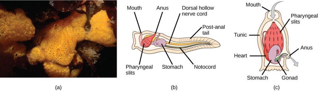
Adult
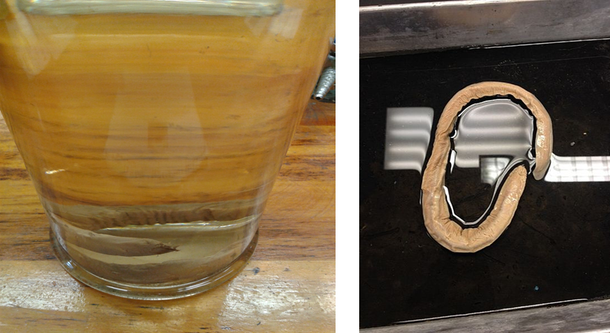
The adult has a thick-walled body sac and an incurrent siphon and excurrent siphon. Gill slits are the only chordate feature retained by the adult form. In some tunicates, the adult form may have been lost. These animals retain the larval form as adults. Examine the preserved tunicate specimens available in the lab.
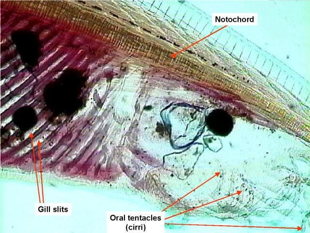
Subphylum Cephalochordata
Members of this subphylum are also called lancelets. Examine a slide of a lancelet using a dissecting microscope and/or compound microscope.
This animal retains all four of the chordate characteristics as an adult. Identify the notochord, nerve chord, pharyngeal gill slits, and post-anal tail.
Notice the segmented pattern of the muscles, also a chordate characteristic.
When feeding, water enters the mouth and moves into the pharynx, a chamber posterior to the mouth. The gill slits are openings in the wall of the pharynx and function to allow water to pass out of the pharynx while filtering particles out of the water. The particles move into the gut for digestion. After passing through the gill slits, water exits via the atriopore.
Examine a preserved lancelet and observe the segmented pattern of muscles and the atriopore located on the ventral surface posterior to the pharynx.
II. Vertebrates
In the previous laboratory, you learned the taxonomy and classification of invertebrate animals. In this laboratory, we will dive deeper into the animal world, and learn about some of the organisms that are vertebrates. Animals that belong to the Subphylum Vertebrata (from the Latin word vertebratus, which means “jointed”), in addition to the notochord, pharyngeal gill slits, a dorsal hollow nerve cord, and a post-anal tail, possess a backbone or vertebral column. Currently, over 69,963 species have been classified in the Subphylum Vertebrata (see ITIS, the Integrated Taxonomic Information System). The reconstruction of the taxonomy, evolution, and phylogenetic relationships within the Subphylum Vertebrata is still an ongoing process. Figure 4 below summarizes the phylogeny of the Phylum Chordata and Subphylum Vertebrata.
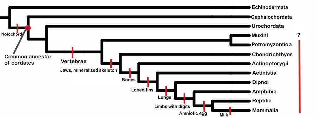
Looking at the phylogenetic tree above, please list all of the anatomical characteristics in each of the vertebrate clades in a table like the one below (the first one is done for you). The traits are indicated on the branches of each new clade.
| Clade Name | Anatomical Characteristic(s) |
| Petromyzontida | Vertebrae |
| Chondrichthyes | |
| Actinopterygii | |
| Actinistia | |
| Dipnoi | |
| Amphibia | |
| Reptilia | |
| Mammalia |
III. Vertebrates Survey
This survey is designed to familiarize you with different clades of vertebrates. You will find an example of animals from each of the various clades.
All vertebrates share common ancestry due to the presence of homologous structures. All vertebrates are deuterostomes. They undergo radial and indeterminate cleavage, have enterocoelous coelom formation, and the anus develops from the blastopore. The table below summarizes eight clades of vertebrates that have representatives alive today (lampreys, cartilaginous fishes, bony fishes, amphibians, reptiles, birds, and mammals). One additional class (not included below) is completely extinct (armored fishes).
Key Terms
| Term | Definition |
| Vertebrate | An animal that has a backbone or spinal column, including mammals, birds, reptiles, amphibians, and fishes |
| Deuterostome | An animal in which anus develops first from the blastopore |
| Fertilization | A fusion of haploid gametes, egg (n) and sperm (n), to form the diploid (2n) zygote |
| Cleavage | Mitotic divisions of a fertilized egg cell |
| Blastula | An animal embryo at the early stage of development when it is a hollow ball of cells |
| Gastrula | An embryo at the stage following the blastula, when it is a hollow cup-shaped structure having three germ layers of cells (ectoderm, mesoderm and ectoderm) |
| Metamorphosis | Additional development that includes significant transformation of an animal after birth or hatching |
| Oviparous | The females lay eggs that hatch outside the mother’s body |
| Ovoviviparous | The female retains the fertilized eggs but does not nourish them, and young are born alive |
| Viviparous | The embryo is nourished via a placenta that develops from the yolk sac of the egg |
| Mammal | A vertebrate animal that has a hair or fur, females secrete milk for the nourishment of the young, and give birth to live offspring (viviparous) |
Pre-lab Activity: Clades of Vertebrates
Using ITIS, the Integrated Taxonomic Information System (see this tutorial on how to search the database), please find an example for each of the clades and complete a table like the one below.
| Clade | Common Name | Example (genus species) |
| Petromyzontida | Jawless fishes | |
| Chondrichthyes | Cartilaginous fishes | |
| Actinopterygii (Osteichthyes) |
Ray-finned bony fishes | |
| Sarcopterygii (Osteichthyes) |
Lobe-finned bony fishes | |
| Amphibia | Amphibians | |
| Reptilia | Reptiles | |
| Aves | Birds | |
| Mammalia | Mammals |
Before coming to the lab, use “Reading: Chordates” (scroll down to the Subphylum Vertebrata—Vertebrates) and your textbook to learn more about animals that are vertebrates.
Clade: Myxini
Example: Hagfish
Read about the habitat, life cycle, and reproduction of the jawless fishes in “Jawless Fishes” and watch the video below to learn more about the morphology and physiological processes of the hagfish.
Morphology
The general characteristics of hagfish are
- Cranium (skull), tongue, thick skin, cartilaginous skeleton, jawless
- Blind, undergo direct development with a larval stage, paired limbs absent
- Notochord present in the adult
Hagfish are long, slender, and pinkish, and are best known for the large quantities of sticky slime that they produce. Hagfish have three accessory hearts, no cerebrum or cerebellum, no jaws or stomach, and will sneeze when their nostrils clog with their own slime. Hagfish are almost blind but have well-developed senses of touch and smell. They have four pairs of sensing tentacles arranged around their mouth. The mouth lacks jaws, but a hagfish is equipped with two pairs of tooth-like rasps on the top of a tongue-like projection. Unlike other fish, the hagfish undergo direct development, with no larval stage. The newly hatched young are practically miniature versions of their parents. Young are hermaphroditic at first, bearing both sets of sex organs. Later in life, they will be either male or female but may change sex from season to season. The reconstruction of taxonomy, evolution, and phylogenetic relationships of the clade Myxini within the Subphylum Vertebrata is still an ongoing process.
Respond
Name three anatomical characteristics of a hagfish mentioned in the video above.
1.
2.
3.
Examine
If there is a preserved hagfish available in the lab, examine it and identify as many of the characteristics (listed above) of this clade as you can.
Clade: Petromyzontida
Example: Lamprey
Read about the habitat, life cycle, and reproduction of the jawless fishes in “Jawless Fishes” and watch the video below to learn more about the morphology and physiological processes of the lamprey.
Morphology
The general characteristics of lamprey are
- Separate sexes (males and females)
- Mate in an anadromous manner
- Vertebral column (vertebra), cranium (skull), tongue, thick skin, jawless
- Mouth with the keratinous teeth
The earliest fish (jawless Agnatha) appeared about 530 million years ago (mya). They evolved from sessile, filter-feeding ancestors in shallow fresh, or brackish water. Their environment was characterized by low salt levels and was at least intermittently oxygen-depleted (hypoxic) (Davenport 2019).
Respond
Why is the lamprey considered to be a parasite?
Name three anatomical characteristics of a lamprey mentioned in the video above.
1.
2.
3.
Examine
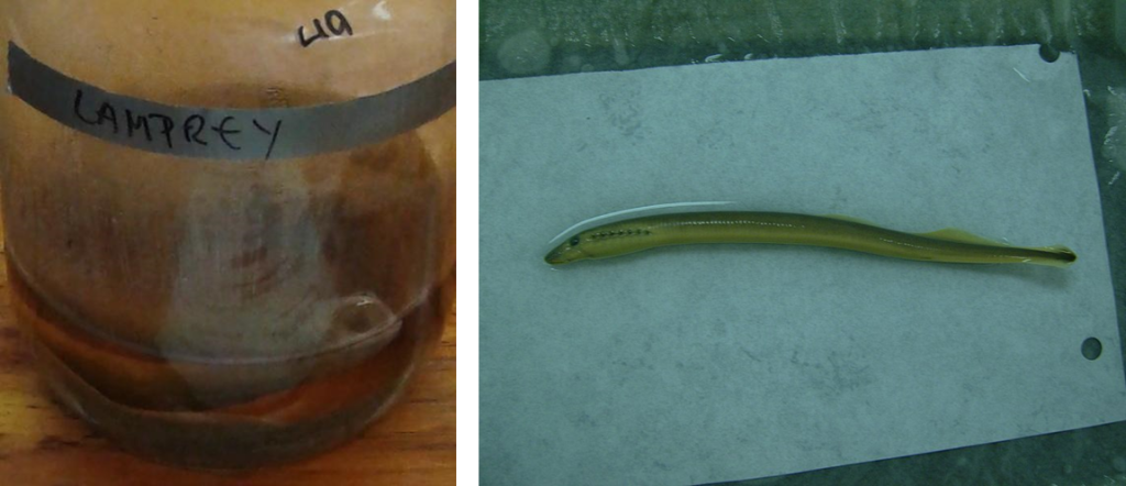
Examine the preserved, dissected lamprey in the lab. Handle it carefully so that it does not get cracked or damaged. Note that the anatomical structures are labeled with small numbers. The name of each structure is on the back of the model. Find the notochord and the vertebral column. What is their position relative to one another?
Observe the cartilaginous skeleton and the lack of jaws. Jaws and bony skeletons evolved later in the vertebrate clade. Together with the hagfish, lampreys are part of the group often called Agnathans (“jawless fish”).
Does the lamprey have teeth? What do lampreys feed on?
Clade: Chondrichthyes
Example: Cartilaginous fishes—shark
Read about the evolution of sharks in “Jawed Fishes” and watch the video below to learn more about the morphology and physiological processes of sharks.
Morphology
The general characteristics of sharks are
- Cartilaginous skeleton
- Teeth not fused to jaws
- No swim bladder
- Intestine with spiral valves
- Placoid scales
- No operculum
- Some ovoviviparous
Respond
Name three anatomical characteristics of Chondrichthyes mentioned in the video above.
1.
2.
3.
Examine
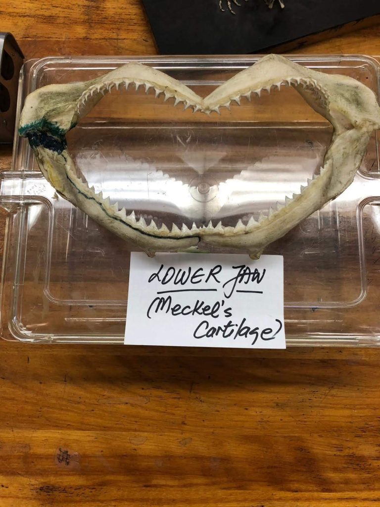
Examine the preserved shark jaws on display. How are the teeth arranged? What do you think the functional reason for this arrangement could be? What are the advantages of this type of dentition?
Examine the preserved cartilaginous shark skeleton. Are there any parts of the skeleton that are mineralized rather than cartilaginous? What are the advantages of a cartilaginous skeleton?
Finally, examine the preserved, dissected shark specimen. Handle it carefully so that it does not get cracked or damaged. Note that the structures are labeled (the names of each structure are on the back of the specimen case). Find the vertebral column. Is there a notochord in the adult, as there was in the lamprey? What happens to the notochord in the adult shark and other cartilaginous fish?
What other adaptations does the shark possess? Can you see the structures that provide a surface for gas exchange? What are they called, and how do their location and shape aid in efficient gas exchange for respiration? What structures does the shark possess for buoyancy? Ask your instructor if you are not sure.
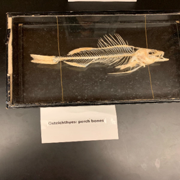
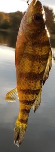
Clade: Osteichthyes (Actinopterygii)
Example: Bony fishes—perch
Read about the habitat, life cycle, and reproduction of the bony fishes in “Jawed Fishes” and watch the video below to learn more about the morphology and physiological processes of bony fishes.
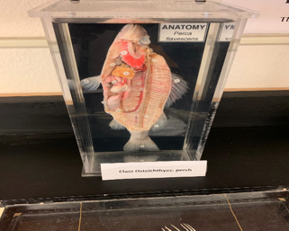
The bony fishes represent the largest group of vertebrates. There are almost 29,000 species of bony fish found in freshwater and marine environments in the world. They are oviparous animals. They have dermal scales, operculum over gill chamber, mineralized endoskeleton, terminal mouth, swim bladder, median and paired fins. Yellow perch is a common freshwater fish in North America.
Respond
What is the function of a swim bladder?
What is the function of the operculum?
Name three anatomical characteristics of a bony fish mentioned in the video above.
1.
2.
3.
Examine
Examine the preserved skeleton of a bony fish. Compare it to the cartilaginous skeleton of the shark. What are the advantages of a bony skeleton?
Examine the preserved, dissected perch on display in the lab. What is the function of the operculum? What advantage does this structure give the bony fish compared to the cartilaginous fish?
Can you locate the swim bladder? What is the evolutionary significance of this trait? In other words, what structure later developed from the swim bladder that influenced the transition of vertebrates onto land?
Clade: Amphibia
Example: Frog
Read about the habitat, life cycle, and reproduction of amphibians in “Amphibians” and to learn more about their morphology and physiological processes.
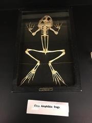
Members of the class Amphibia were the first vertebrates to partially live on land. This superclass includes salamanders, toads, caecilians, and frogs. Frogs are ectothermic vertebrates. They have long modified hind legs and short forelegs. They use their paired appendages to aid in movement on land. The respiratory system is characterized by lungs, gills, and a highly vascularized skin and mouth lining. The center of their circulatory system is a three-chambered heart, unlike the two-chambered heart of fish that you just learned from superclass Osteichthyes above. While the frog’s skeleton is mostly cartilaginous, it consists of a bonier skeletal system (see Figure 13 below). The skin is smooth and moist, containing mucous glands (see Figures 10 and 11 below).
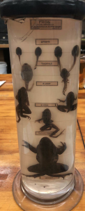
Depending on the frog species it needs water or moist conditions to reproduce. Above all, frogs undergo complete metamorphosis, which means that they start as small tadpoles with a tail in the water and turn into amphibians that can live in water and on land with developed organ systems. Female frogs can lay hundreds of eggs in one clutch. However, most of these eggs will not end up as grown adult frogs. Since frogs deposit their eggs in the water, a large number of these eggs get eaten by fish and other predators. The tadpoles that hatch from surviving fertilized eggs become another essential food source for the organisms in the food chain. Eventually, surviving tadpoles undergo metamorphosis to transform into adult frogs. You will learn more about predator and prey interactions, food webs, and food chains during the next two labs. Figure 9 summarizes the frog life cycle.
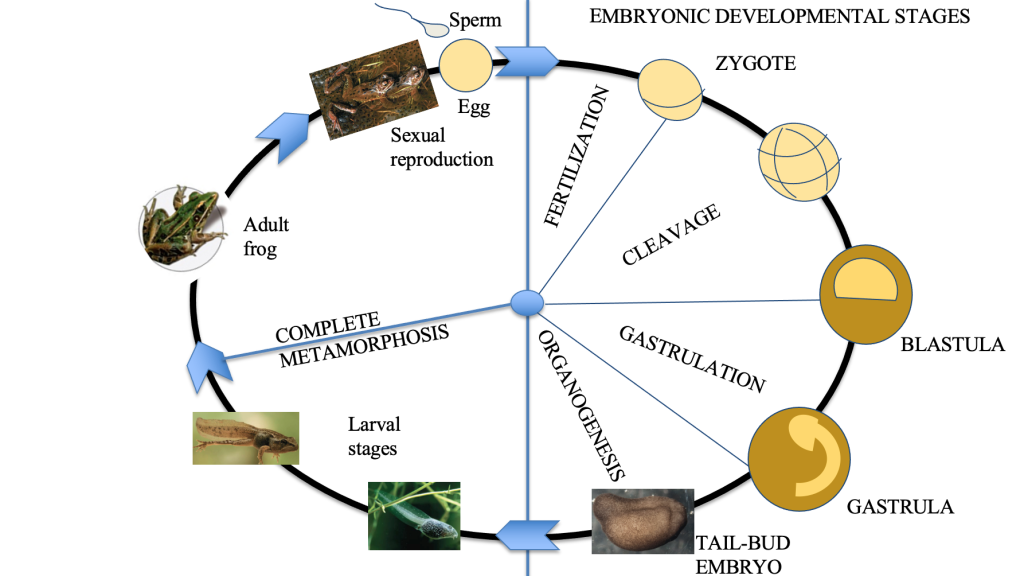
Using your knowledge of an animal life cycle, in one paragraph please describe the life cycle of a frog starting from the adult frog. In your paragraph, refer to Figure 9 above. Also, please use the vocabulary listed in the table of Key Terms, and make sure to indicate whether the transition of each step is accomplished via mitosis or meiosis.
In Activity 1 below, you will examine the external anatomy, oral cavity, digestive, circulatory, reproductive, urogenital, and nervous systems of a leopard frog. You will identify parts of its external and internal anatomies and make comparisons to the human anatomy. Through the dissection experiment, will you be able to conclude whether there are similarities between the organ systems of a frog and the organ systems of a human?
Respond
What is the binomial nomenclature of the leopard frog (genus and species)?
Please, answer whether the frogs are deuterostomes or protostomes.
Activity 1: Leopard Frog Dissection
In this activity, you will examine the external and internal anatomy of the frog. Watch the frog dissection videos below. As you watch, notice where the organs are located and jot down the functions for each structure listed in the chart in Activity 2 below. Then follow the instructions that follow to dissect the frog with your group. Be sure to wear gloves and lab safety goggles, and exercise caution when using sharp instruments.
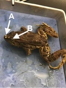
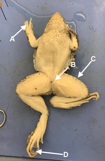
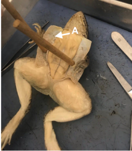
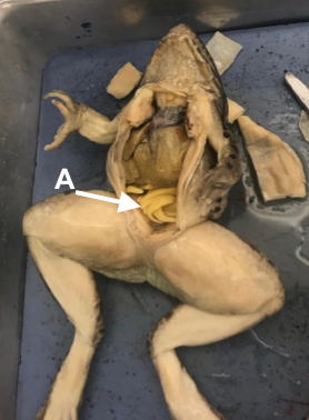
Begin by examining the external anatomy. Follow the instructions and answer the questions below (from the source attributed below). Refer to the photos above. Later on, you will be using the letters on each image to identify the structures indicated.
External anatomy of a leopard frog
- Place the frog in the dissection pan legs down.
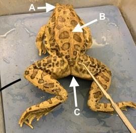
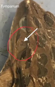
- Identify the eyes, covered by a nictitating membrane, the external nares (nostrils), and the tympanum located behind each eye.
- What is the function of the tympanum?
- Examine the front and back limbs. How many phalanges are on the hindfeet? The forefeet? Which pair of limbs is the longest? How does this assist the frog in its movement?
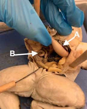
- Mouth (See images below.)
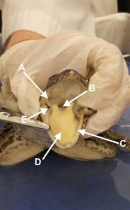
- Turn the frog over and open the mouth as wide as you can. You can cut the hinges of the jaw if necessary. Identify the following structures:
- Two vomerine teeth located in the middle of the roof of the mouth
- Maxillary teeth (smaller) located on the sides of the upper jaw
- Tongue
- Pharynx (located behind the tongue)
- Esophagus, the opening leading to the stomach
- Glottis, slit where air passes through to enter the trachea, which leads to the lungs
- Eustachian tubes (2) openings that lead to the ears. They are located at the angle of the jaw.
Internal anatomy of a leopard frog
In order to continue studying the internal anatomy, we use dissection pins to fasten the frog to the rubbery surface of the dissection pan with the ventral, or front, side up. Using scissors, we can make a superficial cut just left or right of the center from the lower abdomen to the tip of the lower jaw. We use the pins to pin back the skin on both sides to expose the abdominal muscles. We can then lift the muscle layer and cut through the body wall to expose the internal organs.
Now dissect the internal anatomy. Follow the instructions below (from the source attributed below).
Frog body dissection
- Place the frog belly side up in the dissecting tray. You can pin down the limbs if necessary.
- Lift up the skin with forceps midway between the hind legs of the frog. Use scissors to cut the skin along the midline of the frog starting between the hind legs and ending at the neck. Be careful not to cut too deeply.
- Cut the skin horizontally above the hind legs and below the front legs creating skin flaps.
- Pick up a skin flap with forceps and use a scalpel to separate the skin from the muscle below.
- Pin the skin flaps to the dissection tray.
- Repeat the same procedure to cut through the muscles. Create one long incision along the midline of the frog from between the hind legs to the neck. Be careful not to cut too deeply and damage the internal organs. When you reach the area just below the front legs of the frog, turn your scissors sideways to cut through the chest bones and avoid damaging the heart and lungs. Then make horizontal incisions above the rear legs and between the front legs. Use forceps and a scalpel to separate the muscle from the tissue below. Then pin the muscle to the dissection tray.
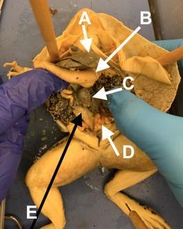
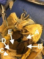
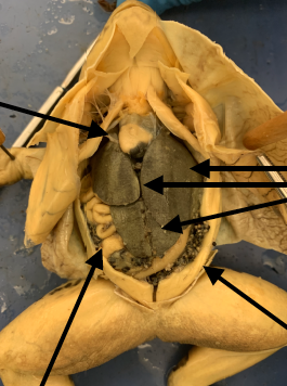
Frog internal anatomy
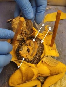
- The most prominent organ is the liver, dark brown in color, and takes up most of the abdominal cavity. Make sure to identify the liver.
- Next, identify the lungs, two small pouches on opposite sides of the frog midline. They may be partially hidden by the liver.
- Lift up the liver and underneath locate the gallbladder.
- Identify the heart covered by the protective pericardium. Frogs have a three-chambered heart with two atria and one ventricle. Try to locate these different areas of the frog heart. How is it a disadvantage to have a three-chambered heart?
- The stomach is a j-shaped organ located underneath the left lobe of the liver. It connects to the esophagus bringing food from the mouth and the small intestine used for nutrient absorption.
- The small intestine connects to the large intestine which carries any undigested material to the cloaca. Frogs have one opening to the outside environment and the cloaca receives materials from the intestine, the urinary system, and the reproductive system.
- Find the pancreas, a yellow ribbon located between the stomach and the small intestine.
- Locate the spleen, shaped similarly to a pea and located near the stomach.
- You will be able to see the yellow, finger-like, fat bodies, which the frog uses to store fat.
- The kidneys of the frog are long and narrow and located along the back body wall.
- Try to find the mesonephric ducts, thin white tubes that carry urine from the kidney to the cloaca.
- If your frog is female, the abdominal cavity will be filled with black and white eggs. The eggs are stored in the ovaries.
- If you have a male frog, locate the testes. The testes are shaped like a bean and located at the top of the kidneys. They are yellow/tan in color.
When you are finished, rinse and pat dry all the dissecting instruments and return them to where you got them. Place the frog and any organs or tissues you removed from it into the plastic bag it came from. Dispose of this in the regular trash. Then rinse the dissecting pan with tap water and return it to where you got it. Finally, throw away your gloves and wash your hands with soap and water. Use the disinfectant spray and paper towels to clean the lab bench where you were working. Then answer the questions below.
Define the following terms related to the positioning of the frog.
a. Dorsal
b. Ventral
c. Anterior
d. Posterior
Without the presence of eggs, how could you tell whether your frog is male or female?
Activity 2: Frog Anatomy
Using information from the above readings and videos, Figures 10 and 13, and the frog dissection, complete a chart like the one below in your lab report. In the middle column, use the identify by figure number and letter where each organ is shown in the figures above; see the example in the first row.
| Organ | Figure | Function |
| Eyes | 10c A | |
| Stomach | ||
| Tympanic membrane | ||
| Liver | ||
| Gullet | ||
| Oviduct | ||
| Glottis | ||
| Teeth | ||
| Intestine | ||
| Eustachian tube | ||
| Egg mass | ||
| Fat bodies | ||
| Heart | ||
| Kidney | ||
| Cloaca |
Before moving on to the reptiles, examine the other amphibian on display. This is a mud puppy (Necturus), and there is a skeleton and a preserved dissected specimen. How does the mud puppy differ from the frog? How is it the same?
Clade: Reptilia
Example: Lizards
Read about the habitat, life cycle, and reproduction of reptiles in “Introduction to Reptiles” and watch the video below to learn more about their morphology and physiological processes.
Morphology
The most visible difference between reptiles and amphibians is that reptiles are covered in dry scales or scutes, while amphibians have moist skins.
The general characteristics of reptiles are
- Four Limbs (except for those that are limbless, such as snakes)
- Thermoregulation – Regulates body temperature ectothermically
- Upper jaw loosely attached to the skull
- Teeth loosely attached to the jaw
- Territorial
As you examine the reptiles on display, look for evidence of these characteristics. Observe the turtle skeleton and preserved turtle dissection and compare them to the snake skeleton and preserved snake dissection. How are they the same? How are they different?
Respond
Name three anatomical characteristics of Class Reptilia mentioned in the video above.
1.
2.
3.
Clade: Aves
Example: Birds
Clade Aves is part of the Class Reptilia.
Read about the habitat, life cycle, and reproduction of birds in “Introduction to Birds” and watch the video below to learn more about their morphology and physiological processes.
Morphology
The general avian characteristics are
- Adaptations for Flight
- Furcula (Clavicle) – Wishbone (flexible)
- Scapula and coracoid: unite at sternum for support
- Sternum/Keel: attachment of powerful flight (pectoral) muscles
- Digits modified to wings
- Fused vertebrae for support
- Pectoral spindle – Sternum, Keel, Clavicle, and Coracoid
- Pygostyle support tail feathers
- Four-chamber heart
- Amniotic development
- Oviparous
- Membranes of amnion remove waste
- Yolk provides food
- Digestion:
- Crop
- Purpose: partially digest food and regurgitation
- Gizzard
- Esophagus
- Intestines
Respond
Name three anatomical characteristics of Clade Aves mentioned in the video above.
1.
2.
3.
Examine
Examine the display materials for Clade Aves. Compare the skeleton of the bird to the skeletons of the reptiles. How are they the same? How do they differ?
Examine the dissected bird specimen. What unique characteristics can you see? What is their function?
Clade: Mammalia
Example: Human
Read about the habitat, life cycle, and reproduction of birds in “Mammals” and watch the video below starting from 6:02 to learn more about their morphology and physiological processes.
Class Mammalia is characterized by the presence of mammary glands, (from Latin mamma meaning “breast”). Females (and sometimes males) produce milk for feeding (nursing) their young, a neocortex (a region of the brain), and fur or hair. Mammals can be divided into three more groups based on how their offspring develop. These three groups are monotremes, marsupials, and the largest group, placental mammals. Monotremes are mammals that lay eggs. The only monotremes that are alive today are the spiny anteater, or echidna, and the platypus. Marsupials have a specialized pouch for the offspring’s development. And, placental mammals all bear live offspring which are nourished before birth in the mother’s uterus through a specialized embryonic organ attached to the uterine wall, the placenta. The placenta is derived from the same membranes that surround the embryos in the amniote eggs of reptiles, birds, and monotremes.
Respond
Are the monotreme mammals viviparous, ovoviviparous, or oviparous animals? Please explain your answer. (Hint: revisit the Key Terms table.)
Morphology
The general characteristics of mammals are
- Endothermy
- Hair or fur on the body
- Mammary glands
- Four chambered hearts
- Sebaceous (oil secreting glands), sudoriferous (sweat) glands, and scent glands
- Heterodont dentition (different types of teeth)
- Diaphragm
IV. Post-lab Questions
To complete this lab and summarize what you have learned, use information from the readings and frog dissection to complete a chart like the one below. In the chart, list the 11 organ systems and then list the organs found within each organ system.
| Organ system | Organs | Function of the organ system |
Attribution
Sections of this lab are adapted from “9.1: Deuterostome Lab” by Lynette Hauser, licensed CC BY 4.0.

_1.JPG)