Chapter 10: Lateralization and Language
10.1: Lateralization
Almost all mammals are bilaterally symmetrical, with a left half that is more or less a mirror image of the right half. The internal organs, however, often tell a different story. We have a single stomach, liver, and heart, none of which are symmetrical. Even paired organs like the lungs or kidneys, are slightly asymmetrical. The brain can most accurately be thought of as a pair of intimately-connected organs with subtle differences in function.
Typically, information from one half of body is sent to the opposite side of the brain and each side of the brain controls the opposite side of the body. However, our eyes and ears have a more complicated pattern of information transfer. Most of the crossing of the information occurs in the brain stem, in or close to the pons (which means bridge). However, this doesn’t mean that that information stays only in the separate hemispheres. The brain’s two hemispheres are connected by white matter tracts that allow the two halves to communicate. Information received by the right hemisphere is quickly shared with the left and vice versa. The largest interhemispheric white matter tract is the corpus callosum, which is made up of 200-250 million axons. If you held a human brain and separated the two hemispheres dorsally along the longitudinal fissure, you would be able to see the fibers of the corpus callosum holding the two halves together. The corpus callosum is about 10 cm (~4 inches) long from anterior to posterior, and the middle part of the structure forms the dorsal-most roof of the lateral ventricles.
In addition to the corpus callosum, there are a handful of other white matter tracts that allow the hemispheres to communicate. The much-smaller anterior commissure is a tenth of the thickness of the corpus callosum, connects the two temporal lobes, and conveys important limbic information such as memory and emotion. The hippocampal commissure is one of the outputs of the hippocampus that connects the structures in the left and right hemispheres. These small white matter tracts are often used as points of reference in imaging studies or surgical dissection.
In the 1950s, a pair of researchers, Drs. Ronald Myers and Roger Sperry, were very curious about these pathways of communication between the two hemispheres. They wanted to understand how information from one visual field gets conveyed into the opposite hemisphere of the brain. To answer the question of interhemispheric transfer, they conducted a series of experiments in cats and monkeys. As you may remember from Chapter 7, the anatomy of the visual pathways is a a little unusual. Information from each eye is sent to each hemisphere in a very specific way. 50% of the information from each eye crosses to the other side of the brain, so that information about the right visual field from each eye is sent to the left hemisphere and information from the left visual field is sent to the right hemisphere. Sperry and Myers found that when they cut the optic chiasm, which is at the crossing point of the visual pathways – information from each eye was no longer sent to the opposite hemisphere. However, information could still be shared across the hemispheres via the corpus callosum. Cutting both the optic chiasm and the corpus callosum, however, meant that if an animal learned a task with one eye covered, e.g., if the right eye was covered they were unable to demonstrate a similar level of learning when the eye patch was switched to the other eye.
Myers and Sperry then extended their research to humans. Sometimes, cutting the corpus callosum or commissurotomy is suggested for younger patients with drug-resistant epilepsy. Grand mal seizures are often characterized by uncontrolled electrical activity in one hemisphere, which then crosses the corpus callosum to the other hemisphere before “bouncing back” to the original hemisphere. During the procedure, the surgeon cuts the corpus callosum, and in doing so, keeps the atypical electrical activity isolated in one hemisphere. Patients have significantly fewer and less severe seizures following recovery from the operation.
People who have had this surgery are sometimes called split-brain patients, a population of patients who were extensively studied by Dr. Michael Gazzaniga.. Overwhelmingly, split-brain patients are healthy with no significant changes in intelligence and no dramatic changes in personality. However, some of them do experience deficits in memory and concentration.
Among split-brain patients, very unique behavioral and cognitive deficits can be observed under specific experimental circumstances. In a lab setting, we can also use a technique known as hemi-field presentation that capitalizes on the anatomical arrangement of the visual pathways to present information to each hemisphere of the brain separately (Figure 10.1). If we stare at a center spot on a computer screen – information to the right of the spot (i.e., the right visual field of each eye) goes to the left side of the brain and information on the left of the spot (left visual field) goes to the right side of the brain (Figure 10.1). In typical healthy brains, this information is quickly shared across the corpus callosum, but in a split-brain patient, the information stays within each hemisphere. Thus, split brain patients provide scientists with a way to study the function of each hemisphere separately. Myers and Sperry’s human studies noted an interesting difference in the ability of split-brain patients to respond verbally. When the stimulus was sent into the left hemisphere, either using a visual stimulus in the right visual field or an object placed in the right hand, the patients were able to verbalize what they either saw or felt. But, when the stimulus was represented in the right hemisphere, they couldn’t. For example, as in Figure 10.1, if a split brain person looks at a picture with a key in the left visual field (sent to right hemisphere) and a ring in the right visual field (sent to left hemisphere) and are asked what they see, they would say that they only saw a ring because the areas for producing language are typically in the left hemisphere, not the right. However, if they were asked to draw what they saw with their left hand, which is also controlled by the right hemisphere, they would draw the key, but not the ring (Figure 10.1). Myers and Sperry’s conclusion was that the left hemisphere is much better equipped for language-related functions compared to the right hemisphere. Dr. Sperry earned the 1981 Nobel Prize for his work regarding the “effects of disconnecting the cerebral hemispheres”.

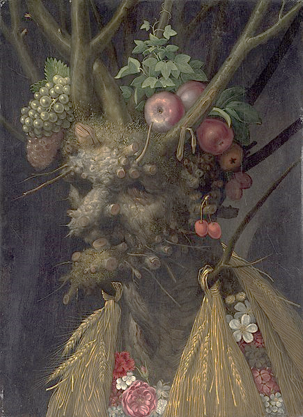
Other studies with split brain patients show that the right hemisphere is better at recognizing faces than the left hemisphere, and is more likely to process information holistically rather than in terms of its individual parts. For example, showing a picture like the one by the artist, Arcimboldo, depicted in Figure 10.2, gives rise to different perceptions depending on which visual field it is shown in. If shown in the left visual field (right hemisphere) split brain patients are likely to perceive a face, but if it is shown in the opposite visual field, they are more likely to perceive the individual elements that the face is made from, such as the fruits and the flowers.

