Chapter 3: Visual Pathways
3.6. Motion Processing: MT and MST
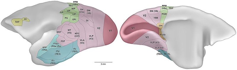
The middle temporal (MT) and middle superior temporal (MST) areas are examples of the specialized visual areas in the brain (see Figure 3.9). Specifically, MT and MST have the function of detecting and processing motion. These motion-selective areas are located just in front of the junction between the temporal lobe and the occipital lobe of the cerebral cortex at the back of the brain. The MT and MST are essential parts of the dorsal stream of the visual cortex, which is responsible for processing spatial location, movement, and object orientation. For example, when catching a football, the MT and MST areas help us with tracking the trajectory of the ball and also help us adjust our movements following the ball’s trajectory.
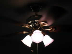
The “motion pathway” is believed to be the stream in which a special role of representing motion signals takes place, beginning with the parasol cells in the retina and continuing through cortical areas of V1 and to MT. MT is known to be the area that receives the input from direction-selective neurons in area V1. Direction-selective means that they have a strong response when an object or field of random dots move in one direction, but they respond little to the other direction. The neurons in MT detect coherent motion in patches. That info from MT is then sent to MST to help put together coherent motion from around the scene to detect optic flow. Evidence for the role of MT and MST in processing motion has been shown through various scientific studies. For example, electrical stimulation in MT can affect our ability to perceive and process visual motion. Damage to these areas can result in a condition called akinetopsia – where a person is unable to perceive movement, such as the spinning of a ceiling fan (Figure 3.10). This makes it tricky to cross the road, drive a car, or even pour a cup of coffee (Purves et al. 2001). Clearly the dorsal stream is important for hand-eye coordination. One brain area that is particularly important for both reaching and grasping is the parietal reach region (PRR) in the parietal cortex (Filimon et al., 2009).
Cheryl Olman PSY 3031 Detailed Outline
Provided by: University of Minnesota
Download for free at http://vision.psych.umn.edu/users/caolman/courses/PSY3031/
License of original source: CC Attribution 4.0
Adapted by: Hannah Thormodsgaard and Matthew Arneson
