Chapter 2: Light and the Eye
2.5 Receptive Fields and Lateral Inhibition
Each cell in the visual system has a receptive field. A visual receptive field is defined as the region of retina in which a change in brightness or color will cause a change in a neuron’s firing rate.
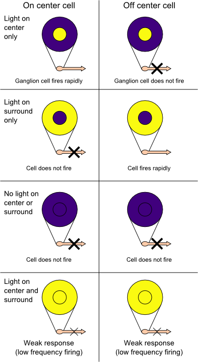
The retinal ganglion cells have receptive fields that have a very basic organization, which resembles two concentric circles. This concentric receptive field structure is usually known as center-surround organization. On-center retinal ganglion cells respond to light spots surrounded by dark backgrounds like a star in a dark sky. If light falls on the entire receptive field, and not just the center, the cell will not increase its firing rate above baseline. Off-center retinal ganglion cells respond to dark spots surrounded by light backgrounds like a fly in a bright sky (see Figure 2.9).
A key function of this receptive field structure is that neurons only respond to edges and objects of a specific size.
These receptive fields with center-surround antagonism are created by a neural process called lateral inhibition. In the retina, lateral inhibition is caused by horizontal and amacrine cells in the retina that integrate responses across the other retinal cells.
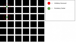
Lateral inhibition has often been used to explain Mach bands, and the gray dots that appear between intersections in the Hermann grid illusion (Figure 2.10). However, there are holes in this explanation — see this explanation from Peter Schiller’s lab at MIT. Lateral inhibition in the retina helps us to see the edges of objects more easily.
Simultaneous contrast
Simultaneous contrast is the visual effect when a gray patch looks lighter when it’s next to a darker patch (Figure 2.11). This shows how fluid our perception of lightness is.

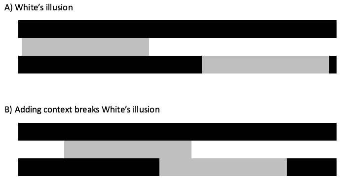
Often, lateral inhibition (phenomenon in which a neuron’s response to a stimulus is inhibited by excitation of a neighboring neuron) is used as an explanation for simultaneous contrast. But White’s illusion (Figure 2.12) shows that this is an inadequate explanation—the rectangles with their long sides against a white background look lighter than the rectangles with their long sides against a dark background. So we know that some other top-down effect is at play in shaping our lightness perception. There are many ways of generating context effects that create lightness illusions. What is important to remember is that our sense of lightness, brightness, and color is easily swayed by context.
Neural responses representing luminance boundaries are more credible than neural responses representing uniform patches for two reasons. First, adaptation makes it impossible to make absolute lightness judgments, so most of our perception is based on contrast and comparisons between two things. Second, the center/surround receptive field structure of our retinal and thalamic visual neurons provides weak responses to uniform fields and strong responses at boundaries. This is why we are very susceptible to lightness illusions: we know that our ability to judge lightness is weak, and we are easily swayed by what we see at boundaries and what we believe about overall scene illumination.
Cheryl Olman PSY 3031 Detailed Outline
Provided by: University of Minnesota
Download for free at http://vision.psych.umn.edu/users/caolman/courses/PSY3031/
License of original source: CC Attribution 4.0Scholarpedia, “Receptive field” by Dr. Jose-Manuel Alonso & Dr. Yao Chen, SUNY State College of Optometry
URL: http://www.scholarpedia.org/article/Receptive_field
License: CC BY-NC-SA 3.0
