"

Chapter 6: Color Vision

Light is not made of different colors—we perceive color by detecting light of different wavelengths. Short wavelength light is perceived as blue, medium wavelengths as green and long wavelengths as red. By mixing these three primary wavelengths in different combinations we are able to match any color. This is referred to as metameric matching. The trichromatic theory of color vision states that we have three different types of cone, each of which is maximally sensitive to one of the three primary wavelengths. Any wavelength that we can see is coded by the relative activity of the three different cone types, which are commonly referred to as blue (short wavelength), green (medium wavelength), and red (long wavelength) cones (Figure 6.5).

A graph is shown with “sensitivity” plotted on the y-axis and “Wavelength” in nanometers plotted along the x-axis with measurements of 400, 500, 600, and 700. Three lines in different colors move from the base to the peak of the y axis, and back to the base. The blue line begins at 400 nm and hits its peak of sensitivity around 455 nanometers, before the sensitivity drops off at roughly the same rate at which it increased, returning to the lowest sensitivity around 530 nm . The green line begins at 400 nm and reaches its peak of sensitivity around 535 nanometers. Its sensitivity then decreases at roughly the same rate at which it increased, returning to the lowest sensitivity around 650 nm. The red line follows the same pattern as the first two, beginning at 400 nm, increasing and decreasing at the same rate, and it hits its height of sensitivity around 580 nanometers. Below this graph is a horizontal bar showing the colors of the visible spectrum.
Figure 6.5. This figure illustrates the different sensitivities for the three cone types

Color Vision Deficiency

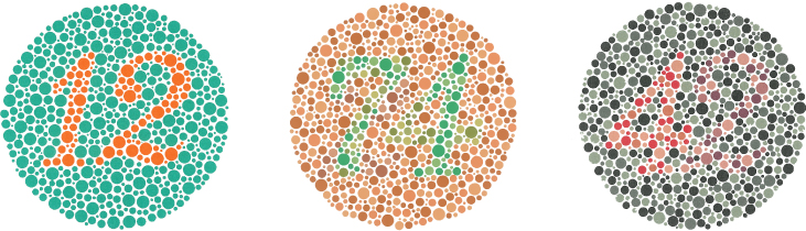
As we already mentioned, most people who are “color-blind” can see colors, but they confuse certain ones because they look similar to them. The psychological term for this condition is color vision deficiency. The circles in Figure 6.6 are part of a common test used to see whether people are able to tell red from green.


Three circles made of dots of different colors. There is a number in each of the three circles
Figure 6.6. The Ishihara test evaluates color perception by assessing whether individuals can discern numbers that appear in a circle of dots of varying colors and sizes. Can you see a number in each of these circles?

Despite much recent improvement in gene therapy for color blindness, there is currently no FDA approved treatment for any form of CVD (color vision deficiency) , and otherwise no cure for CVD currently exists. Management of the condition by using lenses to alleviate symptoms or smartphone apps to aid with daily tasks is possible.

The most common color vision deficiency is confusion between reds and greens due to the absence or malfunction of the green or red cone types. Deuteranopia is the term used when the green cones are not working at all (or are missing), whereas deuteranomaly is a milder form of green cone malfunction. Similarly, protanopia is the more severe form of red cone malfunction and protanomaly is the milder version. Deuteranomaly is the most common type of color vision deficiency. Red-green color deficiency affects males more than females (Birch, 2012). Among males, it affects approximately 8% of European Whites, 5% of Asians, 4% of Africans, and less than 2% of indigenous Americans, Australians, and Polynesians (Birch, 2012). Only about 0.4% in females of European Caucasian descent have red-green color deficiency (Birch, 2012). Males are affected more than females because this is a sex-linked recessive genetic trait. Males inherit this gene from their mothers via the X chromosome. The Y chromosome in males is small and so lacks many of the genes that are present on the X chromosome (like the ones associated with color vision). So, if the recessive gene is present on the X chromosome in a male, it will be expressed—giving rise to red-green color deficiency. Typically, females are not affected because they are likely to also have a dominant normal color vision gene on their other X chromosome.

Other, much rarer, forms of color vision deficiency include tritanopia and tritanomaly, which affect the blue cones. Figure 6.7 shows how individuals who are missing their red, green, or blue cones perceive red, yellow, green and blue.

Figure 6.7. Different Types of Color Deficiency. This figure shows how individuals with different types of color deficiencies see colors. (Credit: Dr. Douglas Keene. Provided by: Wikimedia. License: CC-BY SA 4.0)

Although not especially common, color vision can be affected by diabetes, glaucoma, cataracts, and macula degeneration, as well as by certain drugs and exposure to chemicals. Achromatopsia (a total lack of color vision) can occur because of a congenital lack of cones or may develop after a traumatic brain injury.


License

Icon for the Creative Commons Attribution 4.0 International License

Sensation and Perception Copyright © 2025 by Dr. Jill Grose-Fifer; Students of PSY 3031; and Edited by Dr. Cheryl Olman is licensed under a Creative Commons Attribution 4.0 International License, except where otherwise noted.